Normal Tissue Microarray A103
130 likes | 214 Vues
This microarray contains 54 types of normal tissues, each with 2 tissue spots, for research use. The array is formalin-fixed, paraffin-embedded, and individually packed. It includes detailed documentation and QC information.

Normal Tissue Microarray A103
E N D
Presentation Transcript
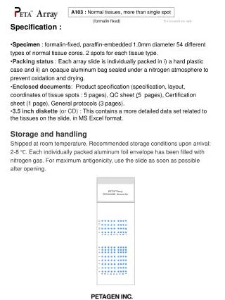
1 2 3 4 5 6 7 8 9 10 11 12 A B C D E F G H I A B C D E F G H I PETATMArray TNV00002P .Section No. A103 : Normal tissues, more than single spot (formalin fixed) For research use only • Specification : • Specimen : formalin-fixed, paraffin-embedded 1.0mm diameter 54 different types of normal tissue cores. 2 spots for each tissue type. • Packing status : Each array slide is individually packed in i) a hard plastic case and ii) an opaque aluminum bag sealed under a nitrogen atmosphere to prevent oxidation and drying. • Enclosed documents: Product specification (specification, layout, coordinates of tissue spots : 5 pages), QC sheet (5 pages), Certification sheet (1 page), General protocols (3 pages). • 3.5 inch diskette (or CD) : This contains a more detailed data set related to the tissues on the slide, in MS Excel format. • Storage and handling • Shipped at room temperature. Recommended storage conditions upon arrival: 2-8 ℃. Each individually packed aluminum foil envelope has been filled with nitrogen gas. For maximum antigenicity, use the slide as soon as possible after opening.
1 2 3 4 5 6 7 8 9 10 11 12 A B C D E F G H I A B C D E F G H I PETATMArray A103 .Section No. 1 2 3 4 5 6 7 8 9 10 11 12 A B C D E F G H I A B C D E F G H I A7 A8 A9 A10 A11 A12 A1 A2 A3 A4 A5 A6 B7 B8 B1 B2 B3 B4 B5 B6 B9 B10 B11 B12 C7 C8 C9 C10 C11 C12 C1 C2 C3 C4 C5 C6 D7 D8 D9 D10 D11 D12 D1 D2 D3 D4 D5 D6 E7 E8 E9 E10 E11 E12 E1 E2 E3 E4 E5 E6 F7 F8 F9 F10 F11 F12 F1 F2 F3 F4 F5 F6 G7 G8 G9 G10 G11 G12 G1 G2 G3 G4 G5 G6 H7 H8 H9 H10 H11 H12 H1 H2 H3 H4 H5 H6 I7 I8 I9 I10 I11 I12 I 1 I 2 I 3 I 4 I 5 I 6 A103 : Normal tissues, more than single spot (formalin fixed) For research use only Layout
A103 : Normal tissues, more than single spot (formalin fixed) For research use only Coordinates of tissue spots
A103 : Normal tissues, more than single spot (formalin fixed) For research use only Coordinates of tissue spots
A103 : Normal tissues, more than single spot (formalin fixed) For research use only Coordinates of tissue spots
A103 : Normal tissues, more than single spot (formalin fixed) For research use only QC sheet_LOT#112120309251 Haematoxylin and Eosin staining A1 A2 A3 A4 A5 A6 A7 A8 A9 A10 A11 A12 B1 B2 B3 B4 B5 B6 B7 B8 B9 B10 B11 B12 C3 C4
A103 : Normal tissues, more than single spot (formalin fixed) For research use only QC sheet_LOT#112120309251 Haematoxylin and Eosin staining C1 C2 C3 C4 C5 C6 C7 C8 C9 C10 C11 C12 D1 D2 D3 D4 D5 D6 D7 D8 D9 D10 D11 D12 E5 E6 E7 E8
A103 : Normal tissues, more than single spot (formalin fixed) For research use only QC sheet_LOT#112120309251 Haematoxylin and Eosin staining E1 E2 E3 E4 E5 E6 E7 E8 E9 E10 E11 E12 F1 F2 F3 F4 F5 F6 F7 F8 F9 F10 F11 F12
A103 : Normal tissues, more than single spot (formalin fixed) For research use only QC sheet_LOT#112120309251 Haematoxylin and Eosin staining G1 G2 G3 G4 G5 G6 G7 G8 G9 G10 G11 G12 H1 H2 H3 H4 H5 H6 H7 H8 H9 H10 H11 H12
A103 : Normal tissues, more than single spot (formalin fixed) For research use only QC sheet_LOT#112120309251 Haematoxylin and Eosin staining I 1 I 2 I 3 I 4 I 5 I 6 I 7 I 8 I 9 I 10 I 11 I 12
Appendix: application protocol 1 Deparffinizing and H&E stain For research use only Deparaffinization and hydration Dry the slide at 58℃ for 1hr or overnight, before deparaffinization (put slides in horizontal position) ① Xylene (removal of paraffin) 4 X 10 min ↓②100% Ethanol (de xylene) 95% Ethanol 1min 95% Ethanol 1min 80% Ethanol 1min 70% Ethanol 1min ↓③ Wash (tap water) until washing is completed (5 min) Routine H&E stain① Wash (tap water) until washing is completed (5 min) ↓ ② Hematoxylin (Harris,nucleus staining,over staining) 3 min ↓ ③ Wash (tap water) ↓ ④ Decolor in 0.1% HCl, 70% Ethanol : repetitive dipping ↓ ⑤ Neutralization 10 min (tap water 5min/ Ammonia water repetitive dipping) ↓ ⑥ Eosin (cytoplasm staining) 1 min ↓ ⑦ Washing 30 sec ↓ ⑧ 70% Ethanol 1 min 80% Ethanol 1 min 95% Ethanol 1 min 95% Ethanol 1 min 100% Ethanol 1 min ↓ ⑨ Xylene (clear to increase refractive index to 1.5 fold) 4 X 10 min ↓ ⑩ Mount with mounting solution ( eg. balsam)
Appendix: application protocol 2 Immunohistochemistry For research use only IHC(immunohistochemistry) Immunohistochemistry is an exquisitely sensitive method for locating an antigen within a cell or tissue through a high-resolution image (a single cell among thousands or millions). The method is based on the use of a primary antibody binding specifically to its cognate antigen. The bound antibody is then visualized by colorimetric or fluorescent detection methods.