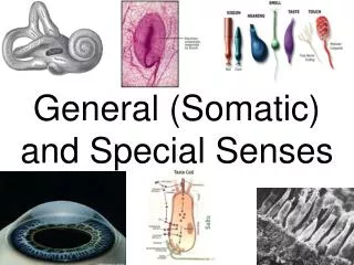
Somatic and Special Senses
570 likes | 891 Vues
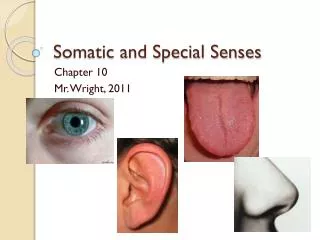
Somatic and Special Senses. Chapter 10 Mr. Wright, 2011. Section 10.1. Introduction. Sensory Receptors. Sensory receptors detect environmental changes and trigger nerve impulses. The CNS then processes and interprets the impulse. Finally, the body reacts to the sensory impulse.

Somatic and Special Senses
E N D
Presentation Transcript
Somatic and Special Senses Chapter 10 Mr. Wright, 2011
Section 10.1 Introduction
Sensory Receptors • Sensory receptors detect environmental changes and trigger nerve impulses. • The CNS then processes and interprets the impulse. • Finally, the body reacts to the sensory impulse.
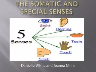
Sensory Receptors • 2 main categories: • Somatic Senses – touch, temperature, and pain • Special Senses – performed by complex sensory organs; things like taste, smell, sight, etc.
Section 10.2 Receptors and Sensations
Types of Receptors • Chemoreceptors – stimulated by changes in chemical concentration • Pain receptors – stimulated by tissue damage • Thermoreceptors – stimulated by changes in temperature • Mechanoreceptors – stimulated by changes in pressure or movement • Photoreceptors – stimulated by light
Sensations • Sensation – a feeling that occurs when the brain interprets a sensory impulse • What you feel is based on which region of the brain receives the impulse. • Projection – your brain’s analysis of where the feeling comes from
Sensory Adaptation • When receptors are continuously stimulated many of them undergo sensory adaptation. • Receptors will stop sending impulses to stop sensation. • Examples: • You don’t “feel” your clothes you are wearing • Garbagemen don’t notice the smell anymore • Water feels hot when you get into a hot tub, but only for a little while.
Section 10.3 Somatic Senses
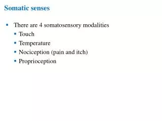
Somatic Senses • Somatic senses - touch, pressure, temperature, and pain.
Touch and Pressure • 3 kinds of receptors: • Sensory Nerve Fibers – free nerves between epithelial cells • Meissner’s Corpuscles – found in hairless portions of skin, respond to motion and light touch. Made of CT. • Pacinian Corpuscles – found deep in skin and in muscles and joints, sense heavy deep pressure. Made of CT.
Temperature • Two types of receptors: warm and cold. • Warm are most sensitive from 77 -113 degrees F. • Cold are most sensitive from 50 – 68 degrees F. • Once you go below 50 or above 113, pain receptors are triggered. • Sensory adaptation does take place!
Pain • Pain receptors are stimulated by tissue damage. • Have little to no sensory adaptation. • A pain receptor may send several impulses… so pain can persist. • How exactly pain receptors are stimulated is poorly understood. • Chemical?
Visceral Pain • Viscera – your guts • Pain receptors in the viscera are the only receptors that produce sensation. • You don’t feel temperature or pressure, but you sure as heck feel pain.
Visceral Pain • Visceral pain may be referred pain. • Referred pain – felt in a different part of the body than the place of stimulation. • For example, people having heart attacks generally feel a sharp pain in their left shoulder. • Caused by common nerve pathways and mistakes by the cerebral cortex.
Types of Pain Fibers • Acute – conduct nerve impulses rapidly, you feel a sharp pain. • Chronic – conduct impulses slowly, you feel a dull, aching pain. • An event that stimulates pain receptors usually stimulates both – first you feel a sharp pain, followed by a dull ache.
Section 10.5 Sense of Smell
Olfactory Receptors • Olfactory – smell • Olfactory receptors are chemoreceptors. • Very closely associated with taste.
Olfactory Organs • Olfactory Organs – yellowish-brown masses in the upper nasal cavity. • Dendrites from nerve cells stick out of epithelium and are covered with cilia. • Odors dissolve into liquids in the nose and bind to these receptors.
Olfactory Nerve Pathways • Stimulated olfactory receptors send impulse to the brain – olfactory bulbs
Olfactory Stimulation • There are many, many different olfactory receptors. • The combination of which receptors are stimulated determines what you smell. • For example: 3, 4, and 8 together might smell like chicken, while 1, 5, and 10 together might smell like chocolate.
Olfaction Lab – Get Ya Stank On, Yo • Create a table like the following on a piece of paper: • You will pick five different objects from around the room (you may use items of your own if you have something). Smell them and fill out the table: • Name of item – what are you sampling? • Describe the smell – what does it smell like? Is it strong? Weak? Does it burn? • Sensory imprinting - sometimes, a smell is tied closely to a certain though or memory. Does the smell bring anything to mind? Does it make you “taste” something in your mouth? • Sensory Adaptation – Give yourself a minute or so to forget the smell. Then, start smelling it again, but this time keep breathing it in. Time how long it takes before you no longer notice the smell.
Section 10.6 Sense of Taste
Sense of Taste • Taste Buds – special organs of taste • Located on the tongue on tiny elevations called papillae. • Also found on the roof and walls of the mouth.
Taste Receptors • Each taste bud contains 50 – 150 taste cells, and each cell is replace every 3 days. • These cells are found within a taste pore, and have small taste hairs that stick out. • Taste hairs are stimulated by liquids in the mouth and send impulse to the brain.
Taste Sensations • 4 primary tastes: • Sweet • Sour • Salty • Bitter • The combination of these tastes that a food brings about is what causes its unique taste. • Pain receptors can also be stimulated.
Section 10.7 Sense of Hearing
The Ear • 3 main parts: • External Ear • Middle Ear • Inner Ear • Functions in hearing and equilibrium.
External Ear • 2 parts: • Auricle – the outer ear (also called the pinna) • External Auditory Meatus – your “earhole” which goes about 2 cm deep • The auricle of the ear “funnels” sound vibrations into the meatus, inside the head.
Middle Ear • Vibrations from the meatus hit the eardrum, causes it to vibrate. • This then causes the auditory ossicles (bones) to vibrate, in order: • Malleus (the hammer) • Incus (the anvil) • Stapes (the stirrups) • The stapes is connected to a membrane called the oval window, causes it to vibrate.
Auditory Tube • There is a small tube that connects the middle ear to the throat – auditory (eustachian) tube • Conducts air into the middle ear to maintain pressure necessary for vibrations. Causes ears to pop!
Inner Ear • We are going to simplify the heck out of this, because it looks something like this and is very complex:
Inner Ear – The Basics • Vibrations hit the oval window. • This causes liquid within the inner ear to start to vibrate. • Vibrations travel through several canals, and will stimulate receptor cells which signal the brain.
Inner Ear – The Basics • Different receptor cells have slightly different sensitivities. • Some sound frequencies will stimulate certain receptors but not others.
Section 10.8 Sense of Equilibrium
Sense of Equilibrium • 2 types of equilibrium – • Static – balance when the head and body are still • Dynamic – balance when the head and body are in movement.
Static Equilibrium • Vestibule – organ of static equilibrium, found between semicircular canals and cochlea • The vestibule is filled with tiny hairs called macula and crystals called otoliths. • As you move your head, the otoliths roll around and press against the macula. • The macula then signal the brain, informing it of the head’s new position.
Dynamic Equilibrium • Organs of dynamic equilibrium – semicircular canals. • Each canal corresponds to a different anatomical plane.
Dynamic Equilibrium • As you move your head in a direction, hairs in the canals move the opposite direction as fluids pass over them. • The combination of these hair’s movements tells the brain how you are moving and about your position.
Dynamic Equilibrium • Other sensory structure also aid with dynamic equilibrium: • Joints in the neck transfer information about position of the body to the brain. • Your eyes also detect changes in posture and signal the brain.
Section 10.9 Sense of Sight –Visual accessories
Accessory Organs - Eyelid • Each eyelid is 4 layers: • Skin (thinnest in the body) • Muscle • Connective Tissue • Conjunctiva – mucus membrane that lines the inner surface of the eyelid, secretes mucus
Accessory Organs – Lacrimal Gland • Lacrimal gland – secretes tears • There are a series of ducts that also carry tears into the nasal cavity. • This is why you might get a runny nose when you cry. • Tears are ALWAYS being produced, even when you’re not crying – they moisten the eye and protect against infection. • Antibacterial
Accessory Organs – Tarsal Glands • The eye also has structures called tarsal glands, which secrete oil to help lubricate the eye.
Accessory Organs - Muscles • There are 6 different muscles that move the eye. • They work together to move certain ways. • OrbicularisOculi controls your blinking.
Section 10.9 Sense of Sight –Structure of the Eye
Structure of the Eye • The eye is a hollow sphere about 2.5cm in diameter. • 3 distinct layers: • Outer Tunic • Middle Tunic • Inner Tunic • Filled with fluids.
Outer Tunic • 2 main parts: • Sclera – • the whites of your eyes, makes up 5/6 of the outer tunic. • Protects the eye and attaches to muscles. • Cornea – • The front 1/6 of the eye, transparent window • Helps focus light into the eye
Middle Tunic • Choroid Coat – posterior 5/6 of the middle tunic, absorbs light and nourishes. • Ciliary Body – anterior 1/6 of the middle tunic, ligaments attach to hold the lens
Middle Tunic • Lens – • Elastic (can change shape) • Allows the eye to focus on an object • Accommodation – the ability of the lens to change shape to focus