Download
1 / 32
330 likes | 465 Vues
Learn about sugar pucker in nucleotide conformations, base pairing in DNA and RNA, Hoogsteen base pairing, and the interaction between base pairs through IR spectroscopy. Understand how bases stack in aqueous solutions and their osmotic pressure effects.

E N D
FCH 532 Lecture 10 Extra credit posted on website, due on Friday (email or typed copy) Chapter 29
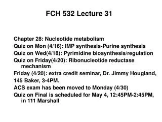
Sugar Pucker • Ribose rings will undergo sugar pucker (become slightly nonplanar). • Most structures show that 4 of the five ring atoms are coplanar, 5th atom is out of the plane in a half-chair conformation. • Endo conformation-if out of plane atom is the same side of the ring as the C5’ • Exo conformation-if out of plane atom is on the opposite side of the ring as the C5’ • Most nucleoside and nucleotide structures (not in double helices) out of plane atom is either C2’ or C3’. • C2’-endo most common; C3’-endo and C3’-exo are also common.
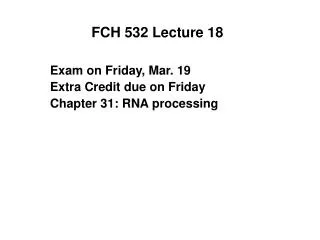
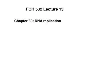
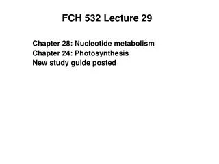
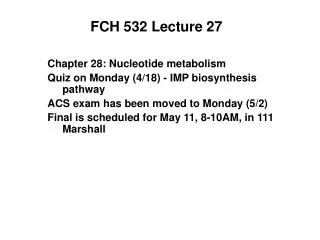
Figure 29-9a Sugar ring pucker. (a) A planar ribose ring (here viewed down the C3—C4 bond) are all eclipsed. Page 1117
Figure 29-9b Sugar ring pucker. (b) The steric strain resulting in Part a is partially relieved by ring puckering in a half-chair conformation in which C3¢ is the out-of-plane atom (same as C5’ so C3’-endo). Page 1117
Sugar Pucker • Endo conformation-if out of plane atom is the same side of the ring as the C5’ • Exo conformation-if out of plane atom is on the opposite side of the ring as the C5’ • Most nucleoside and nucleotide structures (not in double helices) out of plane atom is either C2’ or C3’. • C2’-endo most common; C3’-endo and C3’-exo are also common. • This ribose pucker determines the relative orientatation of the phosphates to the sugar. • To have a regularly repeating model-need C2’-endo (B-DNA) or C3’-endo (A-DNA, RNA-11) • Z-DNA purines are all C3’-endo, pyrimidines C2’-endo (dinucleotide repeating unit). • B-DNA has some flexibility (can be observed as C4’-exo, O4’-endo, C1’-exo and C3’-exo).
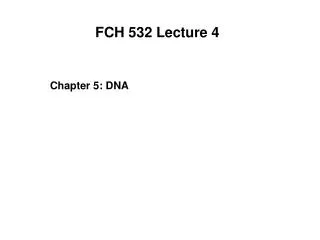
Figure 29-10a Nucleotide sugar conformations. (a) The C3-endo conformation (on the same side of the sugar ring as C5), which occurs in A-RNA and RNA-11. Page 1117
Figure 29-10b Nucleotide sugar conformations. (b) The C2-endo conformation, which occurs in B-DNA. Page 1117
Base Pairing • Watson-Crick pairs most common and only pairing seen in crystal structures of self-complimentary oligonucleotides. • Other geometries available.
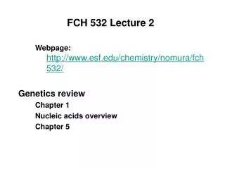
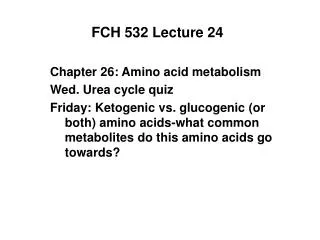
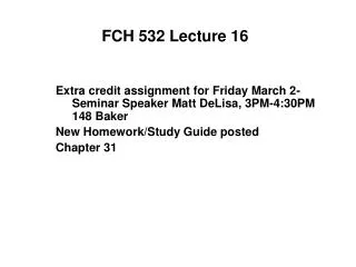
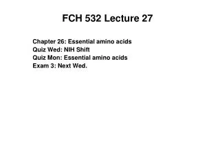
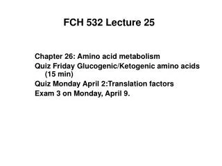
Figure 5-12 Watson-Crick base pairs. Adenine pairs with thymine. Guanine pairs with cytosine. These base pairs are interchangeable in the double helix without altering the positions of the sugar phosphate backbone. The top edge of each base pair is structurally distinct from the bottom edge. The deoxyribose residues are asymmetric. Minor groove exposes the edge from the C1’ atom (open toward bottom) Major groove exposes the opposite edge of each bp. Page 88
Hoogsteen base pairing • Monomeric A and T derivatives form A-T base pairs with adenine N7 as the hydrogen bonding acceptor (normally N1). This is Hoogsteen base pairing • Steric influences make Watson-Crick base pairing the norm for double helices. • Hoogsteen base pairs are biologically relevant. They help stabilize tRNA tertiary structure. • G-C always form Watson-Crick base pairs in crystallized structures because of their 3 hydrogen bonds.
Figure 29-12a Some non-Watson–Crick base pairs.(a) The pairing of adenine residues in the crystal structure of 9-methyladenine. Page 1118
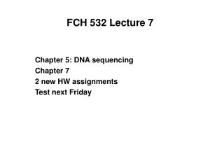
Figure 29-12b Some non-Watson–Crick base pairs. (b) Hoogsteen pairing between adenine and thymine residues in the crystal structure of 9-methyladenine·1-methylthymine. Page 1108
Figure 29-12c Some non-Watson–Crick base pairs. (c) A hypothetical pairing between cytosine and thymine residues. Page 1118
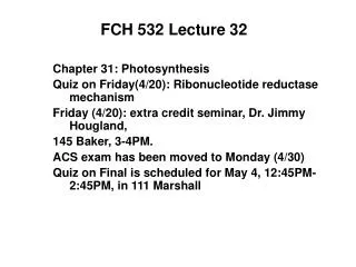
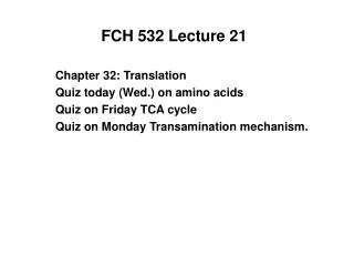
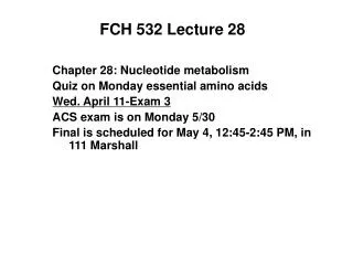
Watson-Crick pairs have high mutual affinity • IR spectroscopy in the N-H region of region of guanine and cytosine derivatives separately and in a mixture shows the interaction between base pairs. • New band near 3500 cm-1 for G+C indicates the interaction. • The same calculated and observed spectra for G+A indicates the lack of interaction. • Can be described by association constant K. [B1 • B2] B1 • B2 B1 + B2 K= [B2] [B1]
Figure 29-13 The IR spectra, in the N—H stretch region, of guanine, cytosine, and adenine derivatives. Page 1119 G+C spectrum
Table 29-2 Association Constants for Base Pair Formation. Page 1119
Figure 29-14 Stacking of adenine rings in the crystal structure of 9-methyladenine. Page 1120
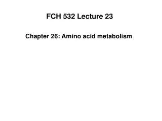
Bases stack in aqueous solution • Bases will aggregate together in aqueous solution. • Interaction can be described by a variation of the van’t Hoff law of osmotic pressure: =RTm • is the osmotic pressure • m=molality of solution (mol solute/kg solvent) • R=gas constant • T=temperature • The molecular mass, M, of an ideal solute can be determined from its osmotic pressure. M=c/m, where c=g solute/kg solvent
Bases stack in aqueous solution For solutions that aggregate, van’t Hoff law is rewritten as = RTm the osmotic coefficient: indicates the solute’s degree of association and varies from 1 (no association) to 0 (infinite association) • The variation of with m for nucleic acid bases in solution is consistent with a model where bases aggregate in a step-wise fashion: A + A A2 + A A3 + A … An Where n is at least 5 (if the reaction goes to completion, 1/n) This result is not based on hydrogen bonding since N6, N6-dimethyladenosine cannot form hydrogen bonds to itself. Aggregation instead results from stacks of planar molecules. Corroborated with NMR studies. Single-stranded polynucleotides show a similar trend.
Figure 29-15 Variation of the osmotic coefficient with the molal concentrations m of adenosine derivatives in H2O. No association Page 1121 ∞ association molality of solution (mol solute/kg solvent)
Figure 29-16 Melting curves for poly(A) and ApA. Page 1121 Broad increase in UV abs with increase in temp for poly(A) and dinucleotides
Table 29-3 Thermodynamic Parameters for the Reaction. Page 1121
Nucleic acids stabilized by hydrophobic forces • Stacking interactions in aqueous solutions are mostly stabilized by hydrophobic forces. • Base stacking is enthalpically driven and entropically opposed. • Hydrophobic intereactions responsible for stability of base stacking nucleic acids are diametrically opposed in character to those that stabilize proteins (enthalpically driven but entropically opposed)
Ionic interactions • Electrostatic interactions are also important. • Melting temperature of double-stranded DNA increases with the cation concentration since these ions bind more tightly to duplex DNA(higher anionic charge) than to single-stranded DNA. • Increased salt shifts equilibrium toward duplex DNA. • Tm = 41.1XG+C + 16.6 log[Na+] +81.5 • XG+C = mole fractionof G+C content and is valid if : 0.3< 1XG+C <0.7 and 10-3 M< [Na+]< 1.0 M Can be used for other monovalent cations (K+, Li+) Divalent cations such as Mg2+, Mn2+, Co2+ bind specifically to phophate groups and are more effective shielding agents. 1 Mg2+ ion is the equivalent of 100 to 1000 Na+ ions.
Comparison of open and supercoiled DNA molecules by electron microscopy supercoiled The two circular molecules have the same mass but very different physical properties RG Open circle RG RG (open circle) > RG(supercoiled) This difference ccounts for the greater mobility of supercoiled DNA during electrophoresis and centrifugation
DNA topology Relative radius of gyration Linear>circles>supercoils • The same double-stranded DNA molecule can have different conformations Frictional coefficient influences electrophoretic mobility and sedimentation rate. Circular Linear “open” Supercoiled
Detecting different DNA topoisomers A. Electrophoretic mobility B. Sedimentation rate Which form goes fastest in each technique?
Different topoisomers of circular DNA can be separated by electrophoresis Supercoiled molecules with increasing twists
Detecting different DNA topoisomers by intercalation of dye molecules
Detecting different DNA topoisomers Gel electrophoresis of a mixture of linear, open, and supercoiled DNA treated with EB A C Sedimentation rate Top (- electrotrode) To t2hrs B Linear/open (Ethidium bromide concentration) Supercoiled>open>supercoiled (negative) (______) topoisomers Bottom (+ electrotrode)
Tertiary structure in DNA • Supercoils refer to the DNA structure in which double-stranded circular DNA twists around each other. Supercoiled DNA contrasts relaxed DNA; • In DNA replication, the two strands of DNA have to be separated, which leads either to overwinding of surrounding regions of DNA or to supercoiling; • A specialized set of enzymes (gyrase, topoisomerases) is present to introduce supercoils that favor strand separation; • The degree of supercoils can be quantitatively described.
Varieties of supercoiled DNA Toroidal spirals within supercoil