FCH 532 Lecture 2
FCH 532 Lecture 2. Webpage: http://www.esf.edu/chemistry/nomura/fch532/ Genetics review Chapter 1 Nucleic acids overview Chapter 5. Bacterial genetics. Advantages can be grown quickly (20 min doubling times). Bacteria usually haploid- phenotype indicates genotype (usually).

FCH 532 Lecture 2
E N D
Presentation Transcript
FCH 532 Lecture 2 Webpage: http://www.esf.edu/chemistry/nomura/fch532/ Genetics review Chapter 1 Nucleic acids overview Chapter 5
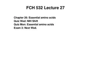
Bacterial genetics • Advantages can be grown quickly (20 min doubling times). • Bacteria usually haploid-phenotype indicates genotype (usually). • Bacterial genetics started in the 1940s for procedures to isolate mutants. • Mutants can be detected and selected for by their ability or inability to grow under certain conditions. • Example: wild-type E. coli can grow on medium with glucose as the sole carbon source. However mutants unable to synthesize leucine require its presence in the growth medium. • Mutants that are resistant to an antibiotic can grow whereas wild-type cells cannot. • Some mutants have proteins that are temperature sensitive. • Use replica plating to screen for colonies with mutant attributes.
Figure 1-30 Replica plating. Page 26
Viruses • Viruses are infectious particles consisting of a nucleic acid molecule enclosed by a capsid (protective coat made of protein). • A virus specifically adsorbs to a susceptible cell and injects its nucleic acid. • The viral chromosome takes over the cell to produce new viruses. • At the end the viral infection causes the lysis of the cell to release the viruses. • Not living organisms since in the absence of their host, they are biologically inert.
Viral complementation and recombination • Bacteriophages-bacterial viruses aka phages • Form plaques (clear spots) on a “lawn” of bacteria. • A mutant phage is detected by its ability to produce progeny under “permissive conditions” and inability to produce progeny under “restrictive conditions”
Viral complementation and recombination • If two different mutant varieties of phage are used to infect a bacterium, it could yield progeny under conditions under which neither of the mutants alone could reproduce through complementation. • Each mutant phage supplies a function that the other mutant cannot. • Each mutation is part of a different complementation group. • Also undergo recombination but differently than eukaryotes.
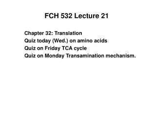
Viral complementation and recombination • Bacteriophages reproduce so rapidly that you can detect recombination with a frequency of 1 in 108. • Benzer carried out studies of the rII region of bacteriophage T4 chromosome. • 4000 bp, approx. 2% of the T4 chromosome. • 2 complementation groups rIIA and rIIB (rapid lysis genes). • In a permissive host (E. coli B) a mutation that inactivates the product of either gene causes the formation of plaques that are larger than wild-type. • In a restrictive host (E. coli K12()), only the wild-type phage can cause lysis. • However, if K12() is infected with 2 different rII mutants, recombination can take place within the same gene.
Figure 1-33 Viral recombination. Page 28
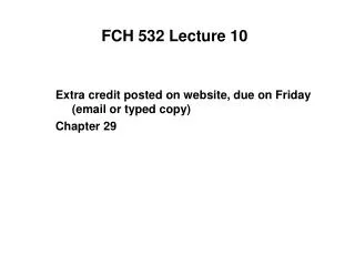
Viral complementation and recombination • Benzer also demonstrated that complementation between 2 mutants in the same complementation group yields progeny in the restrictive host when the 2 mutation are in the cis configuration (on the same chromosome) but do not if in the trans configuration (on physically different chromosomes) • This is due to the fact that when both mutations physically occur in the same gene, the other gene will be intact. • Cistron the functional genetic unit of this type of cis-trans test. • Determined the the unit of recombination is about the size of a single base pair!
Figure 1-34 The cis-trans test. Page 28
Nucleotides and nucleic acids • Form monomeric units of nucleic acids-storage and expression of genetic information. • Nucleoside triphosphates (i.e., ATP, GTP) are compounds that store energy from energy releasing pathways (glycolysis, electron transport) and are use to supply energy for energy-requiring reactions in the cell. • Most metabolic pathways are regulated in part by levels of nucleotides such as ATP and ADP. • Nucleotide derivatives like nicotinamide adenine dinucleotide (NADH), flavin adenine nucleotide (FAD) and coenzyme A are required for many reactions. • Have catalytic activities in in enzymelike nucleic acids (ribozymes).
10.1 RNA and DNA Chemical Structures • Nucleic acids are sequence variable, linear polymers of monomeric units called nucleotides. • A nucleotide is composed of three chemical parts: • An aromatic cyclic compound containing C and N atoms: “nitrogenous bases” • A five-carbon carbohydrate (sugar): an aldopentose. • 1,2, or 3 charged phosphate groups.
General structure of a nucleotide showing the three fundamental units: Purine or pyrimidine nitrogenous base. An aldopentose (D-ribose or 2’-deoxy-D-ribose). 1-3 charged phosphate groups.
Figure 10.3 The aldopentoses in RNA and DNA a) b-D-Ribose b) b-D-2-Deoxyribose
Figure 5-1 Chemical structures of (a) ribonucleotides and (b) deoxyribonucleotides. Page 81 Phosphate group can be bonded to the C5’ or C3’ of the pentose. If bound to C5’ it is a 5’-nucleotide, if bound to C3’, it is a 3’-nucleotide. If there is no phosphate group it is a nucleoside.
A nucleoside consists of a purine or pyrimidine base linked to a carbohydrate (ribose or deoxyribose) by an N-glycosidic bond. Two numbering systems (primed ‘ and unprimed) are necessary to distinguish the two rings.
Nucleotides and nucleic acids • In naturally occurring nucleotides and nucleosides, the bond linking the nitrogenous base to the pentose C1’ atom (glycosidic bond) extends from the same side of the ribose ring as the C4’- C5’ bond. • Nucleotide are moderately strong acids. • Nitrogenous bases are planar, aromatic, heterocyclic molecules. • Derivatives of purines or pyrimidines. • Pyrimidines are 6-membered rings similar in structure to benzenes and have N at at the 1 and 3 positions. • Purines are heterocyclic compounds consisting of a pyrimidine attached to an imidazole group.
Figure 10.2 The major and some minor heterocyclic bases in RNA and DNA. All are derived from purine or pyrimidine.
Figure 10.2 The major and some minor heterocyclic bases in RNA and DNA. All are derived from purine or pyrimidine.
Purines and Pyrimidines with Physiological Activity • 5-fluorouracil (Adrucil) - used in cancer treatment • Fluorine atom is very electronegative. • Analog of thymine, inhibitor of DNA synthesis. • Inhibits enzyme thymidylate synthase.
Figure 10.2 The major and some minor heterocyclic bases in RNA and DNA. All are derived from purine or pyrimidine.
Figure 10.2 The major and some minor heterocyclic bases in RNA and DNA. All are derived from purine or pyrimidine. RNA, DNA contains quantities of methylated bases. Methyl groups added by enzymes after incorporation into nucleic acids
Purines and Pyrimidines with Physiological Activity • Trimethylated derivative of purine ring. • Found in plants Coffea arabica, Camellia thea, Cola acuminata and Cola nitida. • Inhibits enzyme phosphodiesterase involved in cell signaling (cAMP).
Purines and Pyrimidines with Physiological Activity • Antivirial agent with trade name of Zovirax. • Used to treat herpes viral infections. • Inhibits enzyme DNA polymerase of herpes simplex. • Activated in cell via phosphorylation of side chain hydroxyl group by kinase enzyme.
Purines and Pyrimidines with Physiological Activity • 6-mercaptopurine (Purinthol) • Blocks synthesis of nucleic acids. • Effective in treatment of leukemia. • Affects rapidly growing cancer cells.
Formation of nucleotides (phosphorylation) • Linkage of nitrogenous base and aldopentose forms nucleoside. • Nucleotide is formed when phosphoryl group is linked to carbohydrate hydroxyl group
Figure 10.6 Structures for three types of nucleotides AMP ADP ATP
Nucleotides and nucleic acids • In naturally occurring nucleotides and nucleosides, the bond linking the nitrogenous base to the pentose C1’ atom (glycosidic bond) extends from the same side of the ribose ring as the C4’- C5’ bond. • Nucleotide are moderately strong acids. • Nitrogenous bases are planar, aromatic, heterocyclic molecules. • Derivatives of purine or pyrimidine.
Phosphodiester bonds linking mononucleotides into nucleic acids. • The phosphodiester bonds are between the 3’ carbon and the 5’ carbon of the second nucleotide. • This gives direction to the nucleic acids!!! • One end has a free 5’ OH • The other end has a free 3’ OH • The 3’,5’ - phosphodiester bonds are highlighted with green
Structure of AZT and DDI drugs used to treat AIDS 3’-azidodeoxythymidine 2’,3’-dideoxyinosine
DNA base composition follows Chargaff’s Rules • DNA has equal numbers of adenine and thymine residues (A = T) • DNA also has equal numbers of guanine and cytosine residues (G = C) • Structural basis for Chargaff’s rules lie in the hydrogen bonds between the bases. G always hydrogen bonds with C and A always forms base pairs with T. • Base composition of a specific organism is characteristic of that organism (independent of tissue type). • DNA composition varies among different organisms. It ranges from 25% to 75% G + C in different species of bacteria. • RNA, when forming duplexes, also follows Chargaff’s rules
Figure 1-16 Double-stranded DNA. • Each DNA base is hydrogen bonded to a base on the opposite strand forming a base pair. • A bonds with T and G bonds with C forming complementary strands. Page 18