Understanding The Linking Number, Twist, and Writhe in DNA Topology
410 likes | 552 Vues
This lecture explores the concepts of linking number (L), twist (T), and writhe (W) in the topology of DNA. The linking number quantifies how many times one DNA strand winds around another, crucial for understanding supercoiling. We analyze circular DNA's properties and show how L, T, and W are interrelated, adhering to the equation L = T + W. The lecture investigates the roles of topoisomerases in altering linking numbers and offers insights into mechanisms regulating DNA topology during processes such as replication and transcription.

Understanding The Linking Number, Twist, and Writhe in DNA Topology
E N D
Presentation Transcript
FCH 532 Lecture 11 Chapter 29: Nucleic Acid Structures
Linking number • The linking number L of DNA, a topological property, determines the degree of supercoiling; • The linking number defines the number of times a strand of DNA winds in the right-handed direction around the helix axis when the axis is constrained to lie in a plane; • If both strands are covalently intact, the linking number cannot change; • For instance, in a circular DNA of 5400 basepairs, the linking number is 5400/10=540, where 10 is the base-pair per turn for type B DNA.
The twist and writhe • Twist T is a measure of the helical winding of the DNA strands around each other. Given that DNA prefers to form B-type helix, the preferred twist = number of basepair/10; • Writhe W is a measure of the coiling of the axis of the double helix. A right-handed coil is assigned a negative number (negative supercoiling) and a left-handed coil is assigned a positive number (positive supercoiling). • Topology theory tells us that the sum of T and W equals to linking number: L=T+W • For example, in the circular DNA of 5400 basepairs, the linking number is 5400/10=540 • If no supercoiling, then W=0, T=L=540; • If positive supercoiling, W=+20, T=L-W=520;
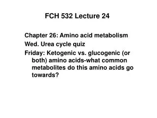
The relation between L, T and W Positive supercoiling
The relation between L, T and W Negative supercoiling
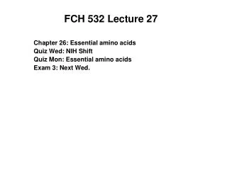
L, T and W calculation • A relaxed circular, double stranded DNA (1600 bps) is in a solution where conditions favor 10 bps per turn. What are the L, T, and W? • During replication, part of this DNA unwinds (200 bps) while the rest of the DNA still favor 10 bps per turn. What are the new L, T, and W? 1400 bps 1600 bps 200 bps L=1600/10=160 W=0 (relaxed) T=L-W =160 L=160 T=(1600-200)/10=140 W=L-T=+20
L, T, and W characterize superhelical DNA • L= linking number = number of times one strand wraps around the other. It is an integer for a closed circular DNA. • T = twists/turns in the DNA ( No. bp/10.4; positive for right-handed DNA • W = writhes =number of turns of the helix around the superhelical axis T = 26 W = 0 L = T + W What kind of number is L??
Figure 29-20 Two ways of introducing one supercoil into a DNA with 10 duplex turns.
Topoisomerases change the linking number of superhelical DNA Type I topos change L in units of one by breaking a single strand of DNA and allowing the duplex to unwind. Type II topos change L in units of two by breaking both strands and allowing a pass-through of both strands of the double helix.
Type I topoisomerases (nicking-closing enzymes) DNA (n turns) + topoisomerase >covalent DNA-enzyme intermediate> dsDNA (n-1 turns) + topoisomerase The formation of a covalent DNA- enzyme complex preserves the free energy of the phosphodiester bond in DNA as a phosphodiester bond between DNA and Tyrosine.
Model of DNA topo I (Ec N-terminus) Note relative size of the enzyme compared to the cross section of the DNA helix, and how the enzyme encircles and holds the DNA. Tyrosine (Y) Position of active site tyr. How does it contact DNA? How do we know it is this amino acid?
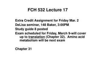
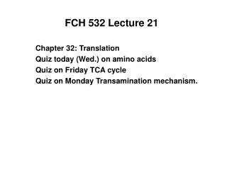
Figure 29-26 X-Ray structure of the Y328F mutant of E. coli topoisomerase III, a type IA topoisomerase, in complex with the single-stranded octanucleotide d(CGCAACTT). Page 1127
Type IA Topoisomerase • Recognize ss region of DNA and gap opening between domains I and II. • DNA cleaved with newly formed 5’end covalently linked to Tyr and the segment with the newly formed 3’ end is noncovalently bound to the protein. • Unbroken strand passed through the opening formed by the cleaved strand to enter protein’s central hole. • Unbroken strand is trapped by the partial closing of the gap • 2 cleaved ends of the cut strand are rejoined in reversal of cleavage reaction • Gap between domains I and III reopens to allow the the rejoined strand to leave the enzyme-the unbroken strand has been passed through the break. • Enzyme returns to original state-If strands formed a negatively supercoiled duplex DNA, L is increased by 1.
Figure 29-27 Proposed mechanism for the strand passage reaction catalyzed by type IA topoisomerases. For (-) supercoiled DNA-L is increased by 1 For circles, they have been catenated or decatenated. Page 1128
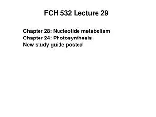
Type IB Topoisomerase • Controlled rotation mechanism • Human topo I is a Type IB enzyme. • Uses Tyr 723 to form phophTyr bond. • Y723F is catalytically inactive mutant, although Y723 would be positioned to nucleophilically attack the P atom of the P-O5’ bond. • Sequence independent binding. • Rotation occurs about sugar-phosphate bonds in uncleaved strand (opposite the cleavage site). • Positive charged amino acids hold the DNA strand in place.
Figure 29-28 X-Ray structure of the N-terminally truncated, Y723F mutant of human topoisomerase I in complex with a 22-bp duplex DNA. Tightly held scissile strand Loosely held strand, free to rotate Page 1129
Figure 29-29 Controlled rotation mechanism for type IB topoisomerses. Binding of supercoiled DNA Formation of noncovalent complex Upstream, cleaved product attached to the enzyme Downstream portion rotates DNA, small rocking motions of enzyme. Covalent intermediate with decreased linking number. Ligation of cleaved strand to intact strand Release of DNA Page 1129
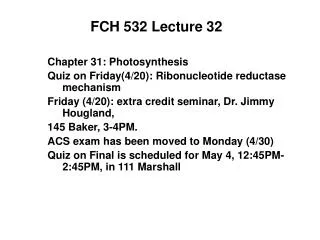
Type II Topoisomerase • Strand passage mechanism • Also known as DNA gyrase • Heterotetramer with A2B2 subunits. A=GyrA, B=GyrB • Prokaryotic Topo II catalyzes the stepwise negative supercoiling of DNA with the concomitant hydrolysis of an ATP to ADP + Pi. • DNA gyrases are inhibited by antibiotics (novobiocin, ciprofloxacin [Cipro]). • Change linking number by 2. • Cuts both strands of duplex, passes the duplex through the break and reseals.
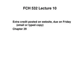
Figure 29-31a Structures of topoisomerase II. (a) X-Ray structure of the 92-kD segment of the yeast topoisomerase II (residues 410–1202) dimer. Page 1131
Proposed mechanism of topo II Arg residues
Type II Topoisomerase • G-segment (Gate segment) binds to enzyme inducing conformational change • ATP binds • T-segment (transported segment) binds causing conformational change that cleaves the G’segment with the A’ subunit. The T-segment is transported through the break in the central hole. • The G-segments are resealed and T-segment is released. • Interface reforms with ATP hydrolysis.
Figure 29-32 Model for the enzymatic mechanism of type II topoisomerases. Page 1131
Prokaryotic Topoisomerase II is a DNA gyrase • In the presence of ATP DNA gyrase can create supercoils; it can relax supercoils in the absence of ATP +AMP, PPi ATP Supercoiled circle Relaxed circle
Figure 29-30A demonstration that DNA gyrase acts by cutting both strands of a duplex, passing the duplex through the break, and resealing it. Page 1130
Inhibitors of DNA gyrase inhibit DNA replication • Two antibiotics, oxolinic acid and novobiocin inhibit replication. E. coli- NovS topoII NovS topoII NovR E. coli- NovS Mutants resistant to novobiocin have a novobiocin-resistant topo II activity in vitro, thus proving that the lethal activity of the drug is its inhiition of DNA topo II activity in vivo. This demonstrates that topo II is an (essential/non- essential) enyme for cell viability.
Summary • DNA exists in different topological forms in vivo and in vitro • DNA topoisomerases catalyze the interconversion of DNA forms • Negative superhelicity (underwinding) helps proteins bind DNA by favoring unwinding of the helix.
Figure 30-1 Action of DNA polymerase. DNA polymerases assemble incoming deoxynucleoside triphosphates on single-stranded DNA templates such that the growing strand is elongated in its 5¢ ® 3¢ direction. Page 1137
Figure 30-2 Autoradiogram and its interpretive drawing of a replicating E. coli chromosome. Page 1137
Figure 11.4 The theta model for replication of a circular DNA model. Note that the DNA structures resemble the Greek letter theta (q). The two replicating forks advance in opposite directions.
Figure 11.3 A photographic image (obtained by autoradiography) of replicating E. coli chromosome. DNA contained 3H-labeled thymidine (b-emitter). Note two replication forks (arrows).
DNA Replication in Bacteria vs. Eukaryotes (mammals) • DNA replication in bacteria involves a single origin of replication site. • 50,000 base pairs/minute DNA synthesis rate in bacteria. • DNA replication in eukaryotes involves multiple ARS elements (autonomously replicating sequence) replication sites. • Only 2,000 base pairs/minute DNA synthesis rate in eukaryotes. • 108 base pairs per chromosome (23 chromosomes) => 1 month to duplicate if only one replication start point.
Figure 11.5. Proposed pathway for replication of eukaryotic DNA. There are several origins of replication (a) A pair of replication forks begins at each origin (b) As the forks advance in opposite directions, the bubbles coalesce to form two double-stranded DNA molecules (c, d, e).
The action of DNA polymerase I Discovered in 1957 by Arthur Kornberg, et al. General reaction catalyzed by DNA polymerase I: dNTP + (dNMP)n (dNMP)n+1 + PPi dNTP = deoxyribonucleoside triphosphates, dATP, dGTP, dCTP, dTTP (dNMP) = preformed DNA with n or n+1 mononucleotides PPi = pyrophosphate
Figure 11.6 The action of DNA polymerase I Preformed DNA performs two roles: One as template (red), which carries the message to be copied. One as a primer (purple) for attachment of added nucleotides.
Figure 10.7 Phosphodiester bonds linking mononucleotides into nucleic acids. • The phosphodiester bonds are between the 3’ carbon and the 5’ carbon of the second nucleotide. • This gives direction to the nucleic acids!!! • One end has a free 5’ OH • The other end has a free 3’ OH • The 3’,5’ - phosphodiester bonds are highlighted with green
Figure 11.6 The action of DNA polymerase I The incoming deoxythymidine triphosphate (dTTP, blue) is held in position by complementary hydrogen bonds to adenine in the template strand. The new phosphoester bond is formed adding a base at the 3’ end of the growing strand. Extra energy is provided by hydrolysis of pyrophosphate.
Table 11.1 Comparison of E. coli DNA polymerases • Primary replicating enzyme in E. coli cells is thought to be DNA pol III: faster, more complex structure. • DNA pol I and II probably serve in editing and repair of DNA.
For the test • Homework problems. • Quiz problems. • You should know key experiments: Griffith, Hershey-Chase, Messelson-Stahl, etc. • Do not need to know codons (table will be provided). • Nucleic acid structures • Supercoiling • Differences in topoisomerases