Download
1 / 37
370 likes | 489 Vues
This lecture covers essential aspects of nucleotide metabolism, focusing on the biosynthesis of nucleotides through key pathways such as IMP and thymine synthesis. It discusses ribonucleotide reductase, its mechanism, and importance in dNTP biosynthesis, alongside the roles of thioredoxin and glutaredoxin in regeneration processes. An overview of nucleotide degradation and the formation of uric acid from purines is also provided, along with information about the upcoming quiz and important exam dates.

E N D
FCH 532 Lecture 27 Chapter 28: Nucleotide metabolism Quiz on Monday (4/18) - IMP biosynthesis pathway ACS exam has been moved to Monday (5/2) Final is scheduled for May 11, 8-10AM, in 111 Marshall
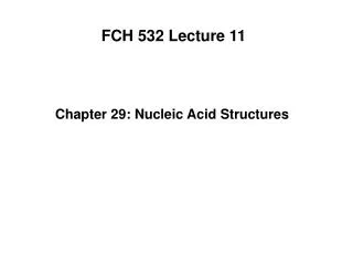
Figure 28-5 Control network for the purine biosynthesis pathway. Feedback inhibition is indicated by red arrows Feedforward activation by green arrows. Page 1075
Overview of dNTP biosynthesis One enzyme, ribonucleotide reductase, reduces all four ribonucleotides to their deoxyribose derivatives. A free radical mechanism is involved in the ribonucleotide reductase reaction. There are three classes of ribonucleotide reductase enzymes in nature: Class I: tyrosine radical, uses NDP Class II: adenosylcobalamin. uses NTPs (cyanobacteria, some bacteria, Euglena). Class III: SAM and Fe-S to generate radical, uses NTPs. (anaerobes and fac. anaerobes).
Proposed reaction mechanism for ribonucleotide reductase Free radical abstracts H from C3’ Acid-catalyzed cleavage of the C2’-OH bond Radical mediates stabilizationof the C2’ cation (unshared electron pair) Radical-cation intermediate is reduced by redox-active sulhydryl pair-deoxynucleotide radical 3’ radical reabstracts the H atom from the protein to restore the enzyme to the radical state.
Thioredoxin and glutaredoxin • Final step in the RNR catalytic cycle is the reduction of disulfide bond to reform the redox-active sulfyhydryl pair). • Thioredoxin-108 residue protein that has redox active Cys (Cys32 and Cys35)-also involved in the Calvin Cycle. • Reduces oxidized RNR and is regenerated via NADPH by thioredoxin reductase. • Glutaredoxin is an 85 residue protein that can also reduce RNR. • Oxidized glutaredoxin is reuced by NADPH using glutredeoxin reductase.
Proposed reaction mechanism for ribonucleotide reductase Free radical abstracts H from C3’ Acid-catalyzed cleavage of the C2’-OH bond Radical mediates stabilizationof the C2’ cation (unshared electron pair) Radical-cation intermediate is reduced by redox-active sulhydryl pair-deoxynucleotide radical 3’ radical reabstracts the H atom from the protein to restore the enzyme to the radical state.
dNTPs made by phosphorylation of dNDP • Reaction is catalyzed by nucleoside diphosphate kinase (same enzyme that phosphorylates NDPs) dNDP + ATP dNTP + ADP • Can use any NTP or dNTP as phosphoryl donor.
Thymine synthesis • 2 main enzymes: dUTP diphosphohydrolase (dUTPase) and thymidylate synthase Reaction 1 • dTMP is made by methylation of dUMP. • dUMP is made by hydrolysis of dUTP via dUTP diphosphohydrolase (dUTPase) dUTP + H2O dUMP+ PPi • Done to minimize the concentration of dUTP-prevents incorporation of uracil into DNA.
Thymine synthesis Reaction 2 • dTMP is made from dUMP by thymidylate synthase (TS). • Uses N5, N10-methylene-THF as methyl donor + dUMP + dTMP
Figure 28-19 Catalytic mechanism of thymidylate synthase. Enzyme Cys thiolate group attacks C6 of dUMP (nucleophile). C5 of the enolate ion attacks the CH2 group of the imium cation of N5, N10-methylene-THF. Enzyme base abstracts the acidic proton at C5, forms methylene group and eliminates THF cofactor Migration of the N6-H atom of THF to the exocyclic methylene group to form a methyl group and displace the Cys thiolate intermediate. Page 1090
F FdUMP 5-flurodeoxyuridylate (FdUMP) • Antitumor agent. • Irreversible inhibitor of TS • Binds like dUMP but in step 3 of the reaction, F cannot be extracted. • Suicide substrate.
Figure 28-20 The X-ray structure of the E. coli thymidylate synthase–FdUMP–THF ternary complex. Page 1091
Thymine synthase oxidizes N5,N10-methyleneTHF • Only enzyme to change the oxidation state of THF. • Regenerated by 2 reactions • DHF is reduced to THF by NADPH by dihydrofolate reductase. • Serine hydroxymethyltransferase transfers the hydroxymethyl group of serine to THF to regenerate N5,N10-methylene-THF and produces glycine.
Figure 28-21 Regeneration of N5,N10-methylenetetrahydrofolate. Page 1091
Nucleotide degradation • Nucleic acids can survive the acid of the stomach • Degraded into nucleotides by pancreatic nucleases and intestinal phosphodiesterases in the duodenum. • Components cannot pass through cell membranes, so they are hydrolyzed to nucleosides. • Nucleosides may be directly absorbed by the intestine or undergo further degradation to free bases and ribose or ribose-1-phosphate by nucleosidases and nucloside phosphorylase. Nucleoside + H2O base + ribose Nucleoside + Pi base + ribose-1-P nucleosidase Nucleoside phosphorylase
Purine nucleoside phosphorylase Catabolism of purines • All pathways lead to formation of uric acid. • Intermediates could be intercepted into salvage pathways. • 1st reaction is the nucleotidase and second is catalyzed by purine nucleoside phosphorylase (PNP) • Ribose-1-phosphate is isomerized by phosphoribomutase to ribose-5-phosphate (precursor to PRPP). Purine nucleoside + Pi Purine base + ribose-1-P • Adenosine and deoxyadenosine are not degraded by PNP but are deaminated by adenosine deaminase (ADA) and AMP deaminase in mammals
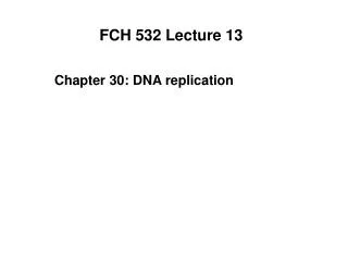
Figure 28-23 Major pathways of purine catabolism in animals. ADA Genetic defects in ADA kill lymphocytes and result in severe combined immunodeficiencey disese (SCID). No ADA results in high levels of dATP that inhibit ribonucleotide reductase-no other dNTPs Page 1093
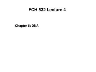
Figure 28-24a Structure and mechanism of adenosine deaminase. (a) A ribbon diagram of murine adenosine deaminase in complex with its transition state analog HDPR. Page 1094
Figure 28-24b (b) The proposed catalytic mechanism of adenosine deaminase. Zn2+ polarized H2O molecule nucleophilically attacks C6 of the adenosine. His is general base catalyst, Glu is general acid, and Asp orients water. Results in tetrahedral intermediate which decomposes by elimination of ammonia. Product is inosine in enol form (assumes dominant keto form upon release from enzyme). Page 1094
Purine nucleotide cycle • Deamination of AMP to IMP combined with synthesis of AMP from IMP results in deaminating Asp to yield fumarate. • Important role in skeletal muscle-increased activity requires increased activity in the citric acid cycle. • Muscle replenishes citric acid cycle intermediates through the purine nucleotide cycle.
Xanthine oxidase • Xanthine oxidse (XO) converts hypoxanthine to xanthine, and xanthine to uric acid. • In mammals, found in the liver and small intestine mucosa • XO is a homodimer with FAD, two [2Fe-2S] clusters and a molybdopterin complex (Mo-pt) that cycles between Mol (VI) and Mol (IV) oxidation states. • Final electron acceptor is O2 which is converted to H2O2 • XO is cleaved into 3 segments. The uncleaved enzyme is known as xanthine dehydrogenase (uses NAD+ as an electron acceptor where XO does not). • XO hydroxylates hypoxanthine at its C2 position and xanthine at the C8 positon to produce uric acid in the enol form.
Figure 28-26a X-Ray structure of xanthine oxidase from cow’s milk in complex with salicylic acid. N-terminal domain is cyan Central domain is gold C-terminal domain is lavender Page 1095
Mechanism for XO • Reaction initiated by attack of enzyme nucleophile on the C8 position of xanthine. • The C8-H atom is eliminated as a hydride ion that combines with Mo (VI) complex, reducing it to Mo (IV). • Water displaces the enzyme nucleophile producing uric acid.
Figure 28-23 Major pathways of purine catabolism in animals. ADA Genetic defects in ADA kill lymphocytes and result in severe combined immunodeficiencey disese (SCID). No ADA results in high levels of dATP that inhibit ribonucleotide reductase-no other dNTPs Page 1093
Purine degredation in other animals Primates, birds, reptiles, insects-final degradation product id uric acid which is excreted in urine. Goal is the conservation of water.
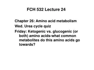
Figure 28-29The Gout, a cartoon by James Gilroy (1799). Page 1097 Gout is a disease characterized by elevated levels of uric acid in body fluids. Caused by deposition of nearly insoluble crystals of sodium urate or uric acid.
Clinical disorders of purine metabolism Excessive accumulation of uric acid: Gout The three defects shown each result in elevated de novo purine biosynthesis
Common treatment for gout: allopurinol Allopurinol is an analogue of hypoxanthine that strongly inhibits xanthine oxidase. Xanthine and hypoxanthine, which are soluble, are accumulated and excreted.
Catabolism of pyrimidines • Animal cells degrade pyrimidines to their component bases. • Happen through dephosphorylation, deamination, and glycosidic bond cleavage. • Uracil and thymine broken down by reduction (vs. oxidation in purine catabolism).
Biosynthesis of of NAD and NADP+ Produced from vitamin precursors Nicotinate and Nicotinamide and from quinolinate, a Trp degradation product Page 1099
Biosynthesis of FMN and FAD from riboflavin FAD is synthesized from riboflavin in a two-reaction pathway. Flavokinase phosphorylates the 5’OH group to give FMN FAD pyrophosphorylase catalyzes the next step (coupling of FMN to ADP). Page 1100
Biosynthesis of CoA from pantothenate Page 1101