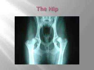
Chapter 20 The Hip
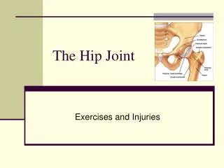
Chapter 20 The Hip. Primary Roles of Hip. Support weight of head, arms, trunk during upright postures and dynamic weight-bearing activities. Provides a pathway for transmission of forces between the lower extremities and pelvis. Anatomy and Kinesiology Osteology and Arthrology. Acetabulum


Chapter 20 The Hip
E N D
Presentation Transcript
Primary Roles of Hip • Support weight of head, arms, trunk during upright postures and dynamic weight-bearing activities. • Provides a pathway for transmission of forces between the lower extremities and pelvis.
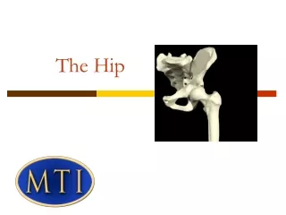
Anatomy and KinesiologyOsteology and Arthrology Acetabulum Fusion of ilium, ischium, and pubis
Anatomy and KinesiologyOsteology and Arthrology Articulation of the femoral head with the acetabular labrum
Two Angular Relationships Angle of inclination of femoral head
Angular Relationships Angle of torsion Projection of the long axis of the femoral head and the transverse axis of femoral condyles
Flexors Iliopsoas TFL Rectus femoris Sartorius Adductor magnus, longus, brevis Pectineus Gracilis Extensors Gluteus maximus Hamstrings Posterior fibers of gluteus medius Piriformis Muscles of the Hip
Abductors Gluteus medius TFL Superior gluteus maximus Gluteus minimus Adductors Adductor group Quadratus femoris Pectineus Obturators Gracilis Medial hamstrings Muscles of the Hip
Medial Rotators TFL Gluteus minimus Anterior fibers of gluteus medius Adductor magnus, longus Semimembranosus/ tendinosis Lateral Rotators Piriformis Obturator interior/exterior Gemelli Quadratus femoris Glut maximus Posterior fibers of gluteus medius Biceps femoris Muscles of the Hip (cont.)
Nerve Supply Lumbar plexus (L1-L4) Sacral plexus (L4-S3) Blood Supply for Head of Femur Artery of ligamentum teres Medium and lateral circumflex arteries Nerve and Blood Supply
Kinematics ROM • Varies with age, sex • Flexion 120–135 degrees with knee flexed 90 degrees • Extension 0–15 degrees • Abduction 0–30 degrees • Rotation generally 45 degrees in each direction (more LR with males, more MR with females)
Kinetics and Kinematics of Gait Single limb stance component of gait
Center Edge Angle – Angle of Wiberg Average adult – 22°–42°
Leg Length Discrepancy (LLD) Unilateral difference in the total length of one leg compared with another. • Anatomic LLD – Actual osseous length difference between the hemipelvis, femur, tibia. • Functional LLD – Position of osseous structures as they relate to each other and to the environment during weight-bearing function.
Examination and Evaluation • History • Lumbar spine clearing examination • Other clearing tests (visceral involvement, knee involvement)
Examination and Evaluation (cont.) • Balance and gait • Joint Mobility and integrity • Muscle performance • Pain and inflammation • Posture and movement • Range of motion and muscle length • Work, community, and leisure integration or reintegration • Special tests
Balance • Balance tests are often included in hip examinations due to high incidence of falls resulting in hip injury: • Berg balance scale • Dynamic gait index • Balance self-perception test • History of balance problems • Type of assistive device used for ambulation
Gait • Gait evaluation is an important component of the examination of a person with a hip dysfunction. • Analysis of gait should include observation of the hip along all three planes of movement during each critical phase of gait. • Of particular importance are the relationships between the hip and the rest of the kinetic chain. • Video analysis can assist in this complex examination procedure.
Joint Mobility and Integrity • Quantity of motion, end feel, and presence/location of pain should be noted during the following tests: • lateral/medial translation • distraction • compression • anteroposterior/posteroanterior glides
Muscle Performance • MMT of hip musculature • Specialized tests looking at positional strength to determine length-associated changes • Selective tissue tension tests to diagnose noncontractile versus contractile lesions • Resisted tests to determine severity of the tissue lesion • Resisted tests can also screen neurologic cause of muscle performance impairment
Pain and Inflammation • Examination is done in conjunction with other tests to determine source (if possible) and cause of pain. • Source diagnosis often requires additional tests that are beyond the scope of physical therapy.
Posture and Movement • Specific lumbopelvic and lower quadrant alignment should be examined about all three planes. • Hypothesis can be developed regarding the contribution of faulty alignments at the ankle, foot, knee, and lumbopelvic regions to the alignment of the hip. • Hypothesis can be generated regarding muscle lengths related to posture alignment. • Initial screening for LLD can be performed.
Range of Motion and Muscle Length • Quick tests: placing foot on standard step, forward bending, squatting, sitting with leg crossed • AROM/PROM in open kinetic chain • Muscle length tests: • Medial/lateral hamstrings • Individual hip flexor lengths • Hip adductors/abductors • Hip rotators
Work, Community, and Leisure Integration or Reintegration • Functional ability can be measured directly through observation of functional tasks. • Self-report measures can also be used. • Harris hip function scale is another self-report measure that is specific to degenerative joint conditions.
Special Tests • Trendelenburg test • Trochanteric prominence angle test (TPAT) • LLD tests • Indirect method – Iliac crest palpation and book correction (ICPBC) • Direct method – Measure distance of fixed bony landmarks using a measuring tape
Impaired Muscle Performance Result of: • Neurologic pathology • Muscle strain • Altered length-tension relationships • General weakness from disuse • Pain and inflammation
Neurologic Pathology • Neuromusculoskeletal or neuromuscular in origin • Neuromusculoskeletal – Pathology at nerve root or peripheral nerve • Treat origin of pathology to positively affect muscle force/torque production
Muscle Strain • Hamstring strains/overuse are common • Treatment focuses on cause of strain • Improving motor control and muscle performance of underused synergists (e.g., gluteus maximus and hip lateral rotators) • Correct biomechanical factors contributing to underused synergists
Muscle Strain • Overstretch can also be a contributing factor to muscle strain. • For example: gluteus medius on high iliac crest side • Strengthen gluteus medius in short range • Taping in short range • Correct posture habits and movement patterns that maintain muscle in lengthened state
Disuse and Deconditioning • Results from injury, pathology, acquired movement patterns contributing to disuse and deconditioning of specific synergists. • Consider acquired postures and movement habits. • Optimize length-associated relationships and restore motor control and force/torque contributions from underused synergists.
ROM, Muscle Length, Joint MobilityHypermobility • Often associated with impairment in the developing hip. • With increasing use of arthroscopy, diagnosis of acetabular labral tears is more common. • Labral tears are a possible precurser to OA
Hypermobility • Hip joint hypermobility has been shown to be associated with OA in numerous studies. • Treatment for developing hip consists of positioning, bracing, or surgery. • Treatment for adult hypermobile hip consists of specific therapeutic exercise, posture education, movement training.
Etiology of Hypermobility • Can be either arthrokinematic or osteokinematic. • Arthrokinematic hypermobility is defined as linear translation that is excessive. • Osteokinematic hypermobility is defined as angular translation that is excessive.
Sahrmann Hip Syndromes Arthrokinematic Hypermobility • Femoral anterior glide syndrome • Femoral lateral glide syndrome Osteokinematic Hypermobility • Femoral adduction with medial rotation syndrome • Femoral adduction syndrome Sahrmann SA. Diagnosis and Treatment of Movement Impairment Syndromes. St. Louis: Mosby, 2002.
Primary Objective of Treatment • Promote joint stability • Prevent continuous stress to overstretched or torn tissues • Posture and movement pattern training • Strengthen lengthened muscles in short range • Improve muscle performance of deep musculature to enhance core stability
Anteversion • Whenever excessive medial rotation ROM is measured, screen for anteversion (TPAT test). • When excessive medial rotation ROM is present, focus on strengthening deep hip LRs. • Educate regarding posture habits and movement patterns.
Functional Approach toTreating Medial Hip Rotation Tendencies
Functional Approach to Treating Medial Hip Rotation Tendencies