Practical training A1 Serum protein electrophoresis Pavla Balínová
Practical training A1 Serum protein electrophoresis Pavla Balínová. Electrophoresis. Cation = positively charged ion, it moves toward the cathode (-) Anion = negatively charged ion, it moves toward the anode (+)

Practical training A1 Serum protein electrophoresis Pavla Balínová
E N D
Presentation Transcript
Practical training A1 Serum protein electrophoresis Pavla Balínová
Electrophoresis • Cation = positively charged ion, it moves toward the cathode (-) • Anion = negatively charged ion, it moves toward the anode (+) • Amphoteric substance = can have a positive/negative/zero charge, it depends on conditions Principle: Some substances have different net charges and can be separated into several fractions in external electric field. But velocity of a particle also depends on the: size, shape of the particle and given applied voltage
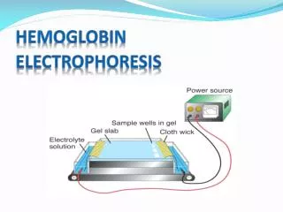
Serum protein electrophoresis on agarose gel • Principle: Serum proteins are negative charged at pH 8.6 (a buffer helps to maintain a constant pH) and they move toward the anode at the rate dependent on their net charge. The separated proteins are fixed and stained by amidoblack solution.
Serum protein electrophoresis on agarose gel is a type of horizontal gel electrophoresis The figure was found at http://www.mun.ca/biology/desmid/brian/BIOL2250/Week_Three/electro4.jpg
Process of electrophoresis • 1. sample application • 2. adjustment ofvoltage or current - DIRECT CURRENT ! (gel-electrophoresis about 70 - 100 volts) • 3.separation time: minutes(e.g. gel-electrophoresis of serum proteins 30 min.) • 4. electrophoresis in supporting medium: fixation, staining and destaining • 5. evaluation: • qualitative (standards) • quantitative (densitometry)
Equipment used for the gel electrophoresisin the practical training A1 power supply (direct current) containers for staining and destaining gel electrophoresis chamber applicator
Serum protein electrophoresisHydragel – agarose gel • Serum proteins are separated into 6 groups: • Albumin • α1 - globulins • α2 - globulins • β1 - globulins • β2 - globulins • γ - globulins Figure is found at http://www.sebia-usa.com/products/proteinBeta.html#
Hydragel 15/30 • Gels with 15 or 30 wells (serum samples) are used in laboratories of clinical biochemistry. • Electrophoresis is also used for separation ofisoenzymes,nucleic acids and immunoglobulins Figure is found at http://www.sebia-usa.com/products/proteinBeta.html#
Hydragel 15/30 • Hypergamma Control Pictured • 16-30 Normal Control Pictured 1-15 Figure is found at http://www.sebia-usa.com/products/proteinControl.html
Evaluation of separated protein fractionsDensitometry • Densitometer is used for scanning of separated proteins in the gel. Scanning the pattern gives a quantitative information about protein fractions. Figure is found at http://www.aafg.org
The use of protein electrophoresis in diagnostics of diseases Electrophoretic patern is constant under physiological conditions(intensity of bands). Spectrum of plasma proteins changes under various diseases(their ratio) evaluation of electrophoretic patern(bands or peaks)
Serum proteins electrophoresis in diagnostics of diseasesNormal pattern Reference ranges: Total protein 6.0 – 8.0 g/dL Albumin 3.5 – 5.0 g/dL α1-globulins 0.1 – 0.4 g/dL α2-globulins 0.4 – 1.3 g/dL β-globulins 0.6 – 1.3 g/dL γ-globulins 0.6 – 1.5 g/dL Figure is found at http://erl.pathology.iupui.edu/LABMED/INDEX.HTM
Acute inflammatory response α1 α2-globulins • Immediate response occurs with stress or inflammation caused by infection, injury or surgical trauma • normal or ↓ albumin • ↑ α1 and α2 globulins Figure is found at http://erl.pathology.iupui.edu/LABMED/INDEX.HTM
Chronic inflammatory response α1 α2 γ-globulins • Late response is correlated with chronic infection (autoimmune diseases, chronic liver disease, chronic infection, cancer) • normal or ↓ albumin • ↑α1 or α2 globulins • ↑↑ γ globulins Figure is found at http://erl.pathology.iupui.edu/LABMED/INDEX.HTM
Liver damage - Cirrhosis γ-globulins • Cirrhosis can be caused by chronic alcohol abuse or viral hepatitis • ↓ albumin • ↓α1, α2 and β globulins • ↑ Ig A in γ-fraction Figure is found at http://erl.pathology.iupui.edu/LABMED/INDEX.HTM
Nephrotic syndrome α2-globulin β-globulin fractions • the kidney damage illustrates the long term loss of lower molecular weight proteins (↓ albumin and IgG – they are filtered in kidney) • retention of higher molecular weight proteins (↑↑ α2-macroglobulin and ↑β-globulin) Figure is found at http://erl.pathology.iupui.edu/LABMED/INDEX.HTM
Monoclonal gammopathy a sharp gamma globulin band • Monoclonal gammapathy is caused by • monoclonal proliferation of β-lymphocytal • clones.These „altered“β-cellsproducean • abnormal immunoglobulin paraprotein. • Production of paraprotein is associated • with benign monoclonal gammopathy • (leucemia) and multiple myeloma. • Paraproteins can be found in a • different position: between α-2 and • γ-fraction. Figure is found at http://erl.pathology.iupui.edu/LABMED/INDEX.HTM