
ELECTROPHORESIS
E N D
Presentation Transcript
ELECTROPHORESIS M.Prasad Naidu MSc Medical Biochemistry, Ph.D,.
ELECTROPHORESIS: • Electrophoresis use the migration of Charged Solutes or particles in liquid medium under the influence of an electric field. • Electrophoresis is widely used analytical technique for the seperation of biological molecules such as plasma proteins, lipo proteins,immunoglobulins,abnormal hemoglobins, genetic variants of transferrin and haptoglobins and isoenzymes
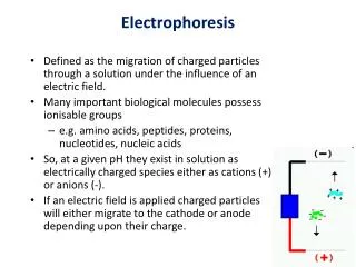
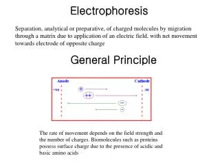
General Principle of electrophoresis: This technique is used for the separation of charged particles. Biological materials such as amino acids, peptides, proteins, nucleic acids posses ionizable groups and hence exist as charged molecules in solutions, either as cations (+vely charged) or anions(-vely charged) depending upon the pH of the medium,.
contd • Even typical nonpolar substance such as carbohydrates can be given charges by derivatization, for example as borate or phosphates. • These charged particles like cations move towards cathode(-vely charged electrode) and anions towards anode (+vely charged electrode) in an electric field.
The difference in Charge: Mass ratio (C/M) forms the basis for the differential migration of particles in an applied electric field. This forms the general principle of electrophoresis. • The Rate of migration of the charged particle depends on • The amount of net charge on the solution • The size and shape of the solute particles • The electrical gradient established in the solution • The pH of the solution Particles of like charges move in an electric field on the basis of their net charge, size, and shape.
Description of technique • Instrumentation and Reagents • A schematic diagram of a conventional electrophoresis system is shown
Made of either platinum or carbon, the polarity of which is determined by the mode of connection to the power supply. • The electrophoresis support • On which separation takes place may contact the buffer directly, or by means of wicks • Two buffer boxes 1. With baffle plates contain the buffer used in the process. Each buffer box contains an electrode. • The entire apparatus is covered • To minimize evaporation and protect the system and is powered by a direct current power supply.
Buffers: • The buffer serves as a multifunctional component in the electrophoretic process as it, • Carriers the applied current. • Establishes the pH at which electrophoresis is performed and • Determines the Electrical charge on the solute.
As the ionic strength of the buffer increases, the proportion of current carried by the buffer will increase and the share of the current carried by the sample will decrease, thus slowing down the rate of migration. So always ionic strength of 0.05M is preferred for the separation of proteins, or lipoproteins in an electric field.
Support Media The Support Medium provides the matrix in which separation takes place. Various types of support media are used in electrophoresis and vary from pure buffer solution in a capillary to insoluble gels (e.g., sheet, slabs, or columns of starch, agarose, or polyacrylamide) or membrances of cellulose acetate.
contd • Gels are cast in a solution of the same buffer to be used in the procedure and may be used in a horizontal or vertical direction. • In either case, maximum resolution is achieved if the sample is applied in very fine starting zone. • Separation is based on difference in charge-to-=mass ratio of the proteins and, depending on the pore size of the medium.
Starch Gel and Cellulose Acetate: Starch gel was the first material to be used as a support medium for electropheresis. Because preparation of a reproducible starch gel is difficult, this medium is now rarely used in the clinical laboratory. Cellulose acetate are seldom used in routine clinical applications. Currently,agarose and polyacrylamide gels are the support media of choice for electrophoresis.
Agarose • Agarose is used in a agarose gel electrophoresis (AGE) for the separation of 1.serum, urine, or cerebrospinal fluid (CSF) proteins, 2. Hemoglobin variants, 3. Isoenzymes, 4.lipoproteins, and 5. Other Substances.
Most routine procedure for AGE are carried out on commercially produced, prepacked microzone gels. Operationally, 0.5 to 1.0 of agarose/DL of buffer provides a gel with suitable strength and good migration properties for proteins and DNA fragments in the range of 0.5 to 20.0 kbp (kilobase pairs)
Polyacrylamide • Polyacrylamide is most useful for mixtures of smaller DNA fragments • The average pore size in a typical 7.5% polyacrylamide gel is about 5nm(50A). • With polyacrylamide, proteins are separated, on the basis of both charge to mass ratio and molecular size, a phenomenon referred to as molecular sieving .
contd • Because of the potential carcinogenic character of acrylamide appropriate caution must be exercised when handling this material if gels are prepared manually
Automated Systems Many laboratories are converting to automated system for electrophoresis, such as the helena SPIFE 3000 or the Sebia Hydragel-Hydrasys system. They are capable of processing of 10 to 100 samples simultaneously. Beckman Coulter Capillary Zone Electrophoresis (CZE) system permits simultaneous processing of seven sample by using multiple capillaries.
contd Newer microchip based analyzers like the Agilent 2100 Bioanalyzer significantly miniaturize and increase the speed of the process for separating proteins, nucleic acids even entire cells.
GENERAL procedure: General Operations performed in conventional electrophoreis include 1. Separation, 2.Staining 3.Detection, and 4. Quantification. Separation: To perform an electrophoretic separation, a hydrated support material such as precast microzone agarose or polyacrylamide, gel is blotted to remove excess buffer and then placed into the electrophoresis chamber.
contd • Care should be taken that the gel has neither excess liquid nor bubbles on it. • Next the sample is added to the support and it is placed in contact with buffer previously added to the electrode Chambers. • Electrophoresis is conducted for a determined length of time under conditions of either constant voltage or constant current
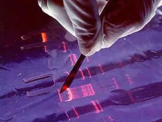
Staining: When electrophoresis is completed, the support is removed from the electrophoresis cell and rapidly dried or placed in a fixative to prevent diffusion of sample components. It is then stained to visualize the individual protein zone. After washing out excess dye, the support is dried.
contd • Stains used to visualize the separated protein fractions are differ according to type of application. • Most commercial methods for serum protein electrophoresis use Amido Black B or members of the Coomassie Brilliant Blue series of dyes for staining. • Silver nitrate or silver diamine has been used to stain proteins and polypeptides.
Detection and Quantification Once electrophoretic separation and staining are complete, it is possible to quantify the individual zones either as a percentage of the total or as absolute concentration by direct densitometry, If the total protein concentration is known.
contd • In the densitometer, the gel (or other medium) is moved past a measuring optical system and the absorbance of each fraction is displayed on a recorder chart or an electronic display
Modern DNA analysis techniques, which may produce several dozen bands of different length DNA fragments require a new type of densitometer referred to as a flat bed scanner or digital image analyzer.
contd • In addition to scanning by densitometry, electrophoresis gels are now being analyzed by mass spectrometers to determine the molecular weights of proteins and their cleavage products, and for peptide sequencing.
Types of Electrophoresis There are mainly two types of Electrophoresis, 1. Vertical Electrophoresis 2. Horizontal Electrophoresis Different types of support media are used in Horizontal Electrophoresis Ex: Filter Paper , cellulose acetate agar gel, agarose gel, starch gel, etc.,
contd • The Vertical Electrophoresismainly use polyacrylamide gel, • Depending upon the nature of supporting medium. • Agar gel Electrophoresis AGE)-where agar gel is used as supporting medium. • Polyacrylamide gel Electrophoresis(PAGE). Acrylamide and methylene bisacry lamide forms a polymer in this case. • Cellulose acetate electrophoresis (Paper Strip Electrophoresis) • Where cellulose acetate paper serves as the supporting medium.
2. Depending upon the mode of the technique: • Slide gel Electrophoresis • Tube gel Electrophoresis • Disc Electrophoresis • Low and high voltage Electrophoresis
Zone Electrophoresis Zone Electrophoresis techniques produce zone of proteins, which are heterogeneous and physically separated from one another as shown . They are classified according to the type and structure of the support material used and are commonly referred to as AGE cellulose acetate electrophoresis (CAE) polyacrylamide gel Electrophoresis (PAGE)etc.,
contd • In 1934, consden introduced a simpler Zone electrophoresis technique using filter paper as a supporting medium . • It uses an inert supporting like paper or gel. It is simple, routinely used and gives better resolution than moving boundary type. • A simple and modified method of moving boundary electrophoresis is the zone electrophoresis.
Various Types of Zone electrophoresis • 1. Paper Electrophoresis • 2. Gel Electrophoresis • 3. Isoelectric Focusing • 4. Cellulose acetate • 5. Immunoelectrophoresis
Free or Moving Boundary Electrophoresis: The Original moving boundary Electrohoresis system was devised by arne Tiselius in 1937. Arne Tiselius was awarded nobel prize in 1948 Tiselius 1937 introduced this technique This consists of a quartz U-shaped tube.
contd • Protein in buffer (pH-8-6) is taken in it and is layered with protein free buffer in both limbs of the tube. • Two electrodes are fitted in the two limbs of cell and joined to the battery. • When electric current is passed through the U-tube, the protein negatively charged migrate towards anode and their rate of migration is
Albumin>α1 Globulin> α2 Globulin>β Globulin>Gamma Globulin When current has flown through for 16 hours front edge of albumin has travelled considerably ahead of other proteins in the order given above. The position of the boundary for each protein can be determined by observing the change in refractive index. The refraction can be photographed in the form of so called electrophoretic pattern
contd • Separation only occurs at the boundaries. • The bulk of the protein remaining homgenous. • It is called moving or free boundary because proteins move freely in solution. • This apparatus is complicated and costly and so was comparatively little used.
Paper Electrophoresis : The technique of paper electrophoresis is simple and inexpensive and requires only micro quantities of plasma for separation. The electrophoresis apparatus in its simplest form consists of two troughs to contain buffer solution, through which electric current is passed.
The plasma under investigation is mixed with bromophenol blue, a blue coloured stain and spotted at the centre of a strip of special filter paper, saturated with an alkaline buffer solution. Barbitone Buffer of pH 8.6 is used. The centre of the filter paper is supported vertically over a glass rod. The ends of the paper dip in buffer solution contained in two troughs placed on either side of the glass rod. The entire system is covered with an airtight transparent lid. .
An electric current of proper amperage and voltage is passed Across the paper, when the charged protein fractions bearing different charges migrate at different rates. The moving boundary of the migrating proteins, stained blue can be seen moving towards anode.
contd • After a run of about five to six hours, the current is stopped and the separated fractions of plasma protein which lie in succession behind the moving boundary, are fixed in their places by heating the paper in an oven at 110 ‘c for half an hour. • The paper is then stained with a solution containing bromophenol blue.
The different fractions appear as blue coloured bands across the filter paper starting from the moving boundary backwards. If a quantitative estimation is required for each fraction, the bands may be carefully cut and eluted, or the bands may be scanned optically in a densitometer. In human plasma five different bands can be identified on paper electrophoresis
GEL ELECTROPHORESIS : This technique involves the separation of molecules based on their size, in addition to the electrical charge. The movement of large olecules is slow in gel electrophoresis. Serum proteins can be separated to about 15 bands, instead of 5 bands on paper electrophoresis. The gels commonly used in gel electrophoresis are agarose and Polyacrylamide, sodium dodecyl sulfate (SDS)
AGAR GEL ELECTROPHORESIS : Slide Preparation About 1.4 ml of Warm (60 °c) agar solution (100 mg / 10 ml of Barbitone buffer) is delivered on a slide uniformly at room temperature and allowed to solidify. Chamber saturation Twenty minutes before starting the experiment, both the buffer tanks of the electrophoretic chamber are filled with equal volume of 0.05 M barbitone uffer (pH8.6) and kept closed to saturate the chamber With solvent vapors.