Electrophoresis
700 likes | 1.19k Vues
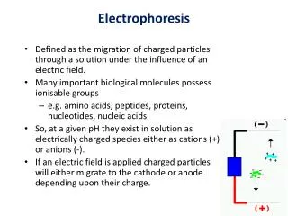
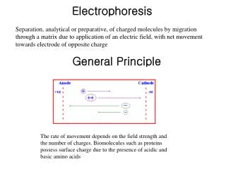
Electrophoresis. Overview. Electrophoresis. Definition refers to the migration of charged solutes or particles in a liquid medium under the influence of an electrical field Iontophoresis limited to the migration of small ions free solution or moving boundary method.

Electrophoresis
E N D
Presentation Transcript
Electrophoresis Overview
Electrophoresis • Definition • refers to the migration of charged solutes or particles in a liquid medium under the influence of an electrical field • Iontophoresis • limited to the migration of small ions • free solution or moving boundary method
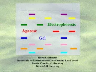
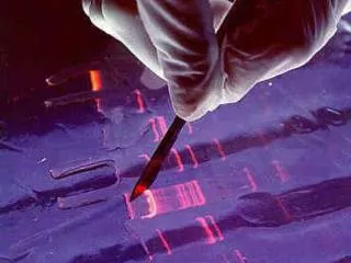
Zone Electrophoresis • Migration of charged molecules • usually in a porous supporting medium • cellulose acetate sheets or agarose gel film • generates an electrophoretogram • Support medium • Protein zones are visualized by • staining with a protein-specific stain
Support medium can be dry and record • Quantitation • Densitometer • Solutes of interest • Proteins in serum, urine, cerebrospinal fluid • Background electrolyte (buffer)
isotachophoresis • applies specifically to the migration of small ions • no background electrolyte (buffer) is mixed with the sample • leading electrolyte solution • contains ions that are faster than any in the sample • Trailing solution • slower than any in the sample • Sample between leading and Trailing electrolyte
isotachophoresis • Applications • separation of small anions and cations, organic and amino acids, peptides, nucleotides, nucleosides, and proteins.
The rate of migration is dependent on • Net electrical charge • Size and shape of the molecule • Electric field strength • Properties of the supporting medium • Temperature of operation
resisting force (counter-force) • Ionic radius of the solute • the viscosity of the buffer solution • electrophoretic mobility • the rate of migration (cm/s) per unit field strength • expressed by the symbol µ • directly proportional to the net charge • inversely proportional to the size of the molecule • inversely proportional to the viscosity of the electrophoresis medium
Factors that can affect mobility • Temperature • Ionic strength • Rate of endosmotic flow • Average pore size of support medium • Point of sample application • wick flow • Drying effect • flow of buffer from both directions
Instrumentation and Reagents • Power Supplies • operation at either constant current • constant voltage • constant power • flow of current through a medium • Resistance, production of heat • Heat • thermal agitation of all dissolved ions • increase in both the migration rate • and the rate of evaporation of water
The water loss • increase in ion concentration • decrease in resistance • increases the conductance of the system • To minimize these effects on the migration rate • it is best to use a constant-current power supply
Pulsed-power or pulsed-field • periodically change the orientation of the applied field • molecules must reorient • new field direction to fit through the pores in gel • reorientation time depends on molecular size • net migration becomes a function of the frequency of field alteration
separation of very large molecules • Such as DNA fragments greater than 50 kilobases
Buffers • they carry the applied current • fix the pH at which electrophoresis is carried out • determine the kind of electrical charge on the solute • the extent of ionization of the solute • they determine the electrode toward which the solute will migrate • The buffer’s ionic strength • concentration of ions • With increasing • molecule becomes more hindered in its movement
Buffers • sharpness of the electrophoretic zones • High ionic strength buffer • reduction in resistance • leads to increased current • excessive heat • leads to denaturation of heat-labile proteins • degradation of other components • sharper band separations • Ionic strength • ion concentration • the charge on the ion
most widely used buffers • barbital buffers • Tris-boric acid-EDTA buffers
Protein Stains • visualize and locate the separated protein fractions • Dyes commonly used in electrophoresis table • Amido Black (Naphthol Blue Black) • Ponceau S • reactivity toward carrier ampholytes • not suitable for polyacrylamide gel-isoelectric focusing • The amount of dye taken up by the sample • type of protein • degree of denaturation of the proteins
Support Media • solutions • such as a sucrose gradient • insoluble gels • sheets, slabs, or columns of starch, agarose, or polyacrylamide,membranes of cellulose acetate
Automated Systems • prepackaged gels, sample application through electrophoresis, staining, scanning of gels, and computation of results. • partially automated • ability to process multiple gels of different compositions • simultaneous processing of seven samples by using multiple capillaries
General Procedures • a hydrated support material • freshly prepared agarose gel • wetted cellulose acetate • Excess buffer removed from the support surface by blotting • bubbles not be present • support is placed in contact with buffer • Sample is applied to the support • and electrophoresis is conducted using either constant voltage or constant current
General Procedures • rapidly dried or placed in a fixative • treated with a dye-fixative reagent (staining) • washing out excess dye • the support is dried • agarose • placed in a clearing agent • cellulose acetate membranes
Detection and Quantitation • Densitornetry • moved past a measuring optical system • the area under each peak • report the results • percentage of each fraction present • in terms of absolute concentration • Reliable quantitation of stained zones • requires light of an appropriate wavelength • a linear response from the instrument • a transparent background
Detection and Quantitation • agarose gels • satisfies the requirement for a clear background • problems associated with densitometry • differences in quantity of stain taken up by individual proteins • differences in protein zone sizes
TYPES OF ELECTROPHORESIS • Starch Gel Electrophoresis • Separates macromolecular ions on the basis • surface charge • molecular size • may be used in a horizontal or vertical direction • proper preparation of gels is relatively difficult • rarely used in the clinical laboratory
TYPES OF ELECTROPHORESIS • Agarose Gel Electrophoresis (AGE) • purified, essentially neutral fraction of agar • a convenient method • applied to the analysis of serum proteins, hemoglobin variants, lactate dehydrogenase and creatinekinaseisoenzymes, lipoprotein fractions • free of ionizable groups • exhibits little endosmosis • exhibits little background staining • native clarity
TYPES OF ELECTROPHORESIS • the agarose surface remain undisturbed • This avoids the surface artifact • Requires sample volume of 0.6 to 3 µL • Electrophoresis time of 30 to 90 min
TYPES OF ELECTROPHORESIS • Cellulose Acetate (CAE) • come as dry, opaque, brittle films • the film is soaked in buffer • Characteristics vary with • extent of acetylation • prewashing procedure used by the manufacturer • the additives used • the pore size • thickness of the membrane
TYPES OF ELECTROPHORESIS • Serum samples (0.3-2.0 µL) are generally applied • may be made transparent • soaking in a solvent mixture • 95 parts methanol and 5 parts glacial acetic acid • Advantage • speed of separation 20 min- 1 h • ability to store for long periods
disadvantage • need for presoaking before use • need clearing the strips prior to densitometry • largely been replaced by agarose gel
TYPES OF ELECTROPHORESIS • Disc Electrophoresis • discontinuities in the electrophoretic matrix • layers of gel that differ in composition and pore size • several proteins with the same electrophoretic mobility • to overcome these deficiencies • Diffusion,broading • Polyacrylamide and Starch gel • pore size is controlled by the percent composition • much smaller than that found in agarose gel
TYPES OF ELECTROPHORESIS • Proteins are separated • on the basis of charge and molecular size • molecular sieving • may yield 20 or more fractions • to study individual proteins in serum • genetic variants and isoenzymes
TYPES OF ELECTROPHORESIS • Polyacrylamide Gel Electrophoresis (PAGE) • three different layers of gel • small-pore separation gel • large-pore gel (spacer gel ) • a large-pore monomer solution contaming a small amount of serum • Separation then takes place in the bottom separation gel • Advantage • Thermostable • Transparent • Strong • relatively chemically inert • can be made in a wide range of pore sizes
potential carcinogenicity • larger pores; less resistance to the passage of large molecules • ideally suited to the separation of DNA fragments up to 20 kilobases • In homogeneous (non-pulsed) electric field
Isoelectric Focusing • The protein migration in a medium possessing a stable pH gradient • moves to a zone in the medium where the pH is equal to the isoelectric point (pI) of the protein. • the charge becomes zero • migration ceases • the protein zones are very sharp • diffusion is also counteracted • acquisition of charge • only 0.02 pH unit
carrier ampholytes • create buffered zones • high concentrations • a high-voltage power source • matrix must be cooled • matrix • polyacrylamide gel • optically clear and supple • large enough pore size • IgM impeded
the anode is surrounded by a dilute acid solution • the cathode by a dilute alkaline solution • Matrix characteristics • Agarose, cellulose acetate • Electroendosmosis-free materials • operating conditions are simple • large pore sizes
Two-Dimensional (2D) Electrophoresis • charge-dependent IEF electrophoresis in the first dimension • molecular weight-dependent electrophoresis in the second dimension • first dimensional in a large-pore medium • Ampholytes are added to yield a pH gradient • The second dimension is often polyacrylamide in a line ar or gradient format
Additives • SDS is used in the second dimension • β-mercaptoethanol in the first • SDS in both dimensions and in sample preparation • Electrophoresis under • Native condition • Denaturing conditions
Detection method • Autoradiography • Using Coomassie dyes • Radiography • fluorographic analysis • method limitation • From 7000 polypeptide spots • Autoradiography • 1100 spots are detected • Using Coomassie dyes • about 400 polypeptides are detected
Coomassie dyes • three times more sensitive than Amido Black • silver staining • 100 times more sensitive than Coomassie dye • Radiography , fluorographic analysis • greatest analytical sensitivity
High-Resolution Electrophoresis • high-ionic-strength buffer • µ = 0.075 • pH 8.6 • admixture of calcium lactate • Support • agarose gel • temperature control is necessary • Serum proteins resolved into as many as 13 zones
High-Resolution Electrophoresis • Discontinuous buffer system • different buffer is used in the electrode chambers from that in the gel • two different pH • alter relative mobility
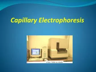
Capillary Electrophoresis • electrophoresis are carried out in capillary tube • 10-100 µm diameter • 20 to 200 cm in length • detector at its terminal end • high-voltage power up to 30 kV • mostly made of fused silica (i.e., pure glass), polyethylene • Advantage • Improved heat dissipation • Sample volumes • in the picoliter to nanoliter range
Capillary Electrophoresis • Reduced separation time • Can be fully automated • applications • low- molecular-weight ions to proteins and other macromolecules • Even uncharged molecules • minimizing band spreading • Improved resolution • Due to narrow bore • a variety of detector types can be used
Capillary Electrophoresis • Detectors • Optical methods • Ultraviolet- visible photometers • Fluorescence • laser-induced fluorescence • Refractive index • Chemiluminescence • Mass spectrometers • Electrochemical detection methods