Download
1 / 42
450 likes | 804 Vues
Radiological aspects of genetic disorders with adult-onset CNS symptoms. Raili Raininko and Atle Melberg. Departments of Radiology and Neuroscience Uppsala University Uppsala, Sweden. Genetic disorders with neurological symptoms: Often pediatric diseases

E N D
Radiological aspectsof genetic disorders withadult-onset CNS symptoms Raili Raininko and Atle Melberg Departments of Radiology and Neuroscience Uppsala University Uppsala, Sweden
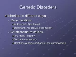
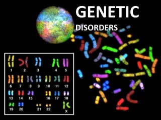
Genetic disorders with neurological symptoms: • Often pediatric diseases • Adult types may occur in some of them • - Disorders giving symptoms only in adulthood
Mitochondrial disorders Clinical onset most often in childhood Course: Static - Progressive - Episodes of exacerbation typical Typical symptoms: epilepsy, progressive external ophthalmoplegia (PEO), myopathy, diabetes mellitus Examples: - Leigh syndrome - MELAS (mitochondrial encephalopathy, lactic acidosis and stroke-like episodes) - POLG-1-associated encephalopathy syndromes
POLG-1 mutation (mitochondrial DNA-polymerase gamma) 1. Autosomal recessive - Epilepsy (episodes of focal status epilepticus) - Stroke-like episodes - Ataxia - Ophtalmoplegia - Sensoric neuropathy - (Liver insufficiency ← Valproate)
POLG-1 mutation 2. Dominant form – a different clinical picture PEO, muscular weakness, parkinsonism, ataxia Diagnosis:- Muscle biopsy - mitochondrial DNA analysis for mtDNA deletions - sequencing POLG-1 gene
POLG-1 mutation, autosomal recessive 18-year-old man CT n:o1 CT 2 days later
POLG-1 mutation 4 days from the symptom onset DWI: trace images b= 1000 T2-w FLAIR
POLG-1 mutation Follow-up: 2 mo 3 wk DWI: trace, b=1000 ADC maps T2-w FLAIR
POLG-1 mutation Patient 2 24-year-old man Twin brother succumbed at age of 19 in status epilepticus T2-w SE
Hepatolenticular degeneration = Wilson´s disease Autosomal recessive Hepatic or neurologic symptoms or dementia Tremor, speech disorders, clumsiness, dystonia, personality changes Defect in copper metabolism Diagnosis: - Blood test: caeruloplasmin, copper - Urine test: copper - Liver biopsy - Genetic analysis, ATP7B gene
Wilson´s disease Man, 34 yr, treated T2-w SE
Polycystic lipomembranous osteodysplasia and sclerosing leukoencephalopathy (PLOSL) = Nasu-Hakola disease : Bone cysts Pain in bones Fractures
Nasu-Hakola disease Later, ~ at age of 30: Symptoms of dementia Death: under age of 50 Autosomal recessive Mutations in DAP12 or TREM2 genes Diagnosis: Clinical + radiological findings
Nasu-Hakola disease Man, 33 yr CT
Nasu-Hakola disease Man 32 yr T2-w SE
Leukodystrophies • - Onset most often in childhood • Most often recessive inheritance • - Also sporadic forms • - Some are x-linked • Uncommon: • Adult-onset forms • Dominant inheritance
Metachromatic leukodystrophy (MLD) Types of MLD: - late infantile - juvenile - adult-onset The adult-onset type most often begins with psychiatric symptoms Diagnosis: Arylsulfatase A activity low in leukocytes Sulfatide excreation ↑ in urine Autosomal recessive different types of mutations in ARSA gene .
Adult-onset metacromatic leukodystrophy Woman, 25 yr Psychiatric symptoms for 6 years T2-w T2-w T1-w PD-w T1-w
Krabbe disease = globoid cell leukodystrophy (GLD) Many types: - congenital - early infantile - late infantile - juvenile - adult-onset Adult-onset type: spastic paraparesis, hemiparesis, ataxia, deteriorating vision Diagnosis: Galactocerebrosidase activity deficiency About 60 different mutations found in GALC gene
Adult-onset Krabbe disease Woman, 28 yr, 5-year history of spastic paraparesis T2-w FSE T2-w FLAIR
Adult-onset Krabbe disease Atrophic spinal cord T2-w FSE
Alexander disease Types: - infantile - juvenile - adult-onset Adult-onset type: diverse symptoms, episodic or progressive course Bulbar and pseudobulbar symptoms, spasticity, ataxia, dementia, nystagmus, palatal myoclonus GFAP mutation, Diagnosis = Genetic tests - Almost all cases sporadic - Autosomal dominant form exists
Adult-onset Alexander disease Woman 34 yr Ataxia, motor clumsiness, exaggerated reflexes, overactive bladder T2-w FLAIR
Adult-onset Alexander disease T2-w FSE Patchy contrast enhancement may occur
Adult-onset Alexander disease C II Pathologic white matter C IV Normal SI Atrophic spinal cord T2-w FSE
Adult onset autosomal dominant leukodystrophy (ADLD) with autonomic symptoms • Age of clinical onset between 40-50 years of age • Autonomic symptoms (bladder and bowel dysfunction, orthostatic hypotension) usually precede cerebellar and pyramidal symptoms • Genetic basis: duplication of Lamin B1 with subsequent overexpression • Diagnosis: - Clinical symptom constellation + MRI - Genetic analysis
Adult-onset ADLD with autonomic symptoms Asymptomatic family member Man, 37 yr
Adult-onset ADLD with autonomic symptoms 34-year-old asymptomatic family member
Adult-onset ADLD with autonomic symptoms Patient, 55 years
Adult-onset ADLD with autonomic symptoms Man, 57 years, with spasticity and ataxia Man, 51 years, wheel chair bound
ControlPatients T2 C 2 C 2 T 6 T 6 T 6 Woman, 59 years Woman, 49 years Man, 51 years
CADASIL= Cerebral autosomal dominant arteriopathy with subcortical infarcts and leukoencephalopathy • Age at onset < 50 yr • At least two of these symptoms: • Stroke-like episodes with permanent neurologic signs • Migraineous headache • Major mood disturbances • Subcortical dementia • Diagnosis: Skin, muscle or brain biopsy • Mutation in the NOTCH3 gene
CADASIL Woman 47 years T2-w FLAIR
Multiple cavernomas Familial form A 21-year-old woman, numbness in the right leg Her brother had had a bleeding in the spinal cord T2-w FSE T2*-w GRE T1-w SE contrast enhanced
Multiple cavernomas Familial form T2*-w GRE SWI sequence
Multiple cavernomas Familial form T2*-w GRE T1-w SE plain contrast enhanced T2-w FSE T2*-w GRE
Hereditary spastic paraparesis and thin corpus callosum (HSP-TCC) • Autosomal recessive • Genetically heterogenic • In 35%, mutation in SPG 11 or SPG 15 gene • SPG11 mutation: central retinal degeneration (Kjellin syndrome) • Spastic paraparesis, cognitive decline, amyotrophy • Symptom onset in 1st to 3rd decade Diagnosis: Clinical constellation + MRI + sequencing of candidate genes SPG 11 and SPG15
Hereditary spastic paraplegia and thin corpus callosum (HSP-TCC) The first MRI at age of 34Female 46 yr.
Thorough and systematic analysis of atrophic and signal intensity changes Ruling out diseases with the same or similar symptoms Specific diagnosis Some alternative diagnoses
Thorough and systematic analysis of atrophic and signal intensity changes Ruling out diseases with the same or similar symptoms Specific diagnosis Some alternative diagnoses Collaboration with clinicians and geneticists essential