Class 1
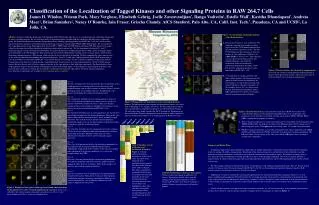
A. Class 1. Class 2. Class 3. Class 4. Class 5. Class 6. Class 7. Classification of the Localization of Tagged Kinases and other Signaling Proteins in RAW 264.7 Cells

Class 1
E N D
Presentation Transcript
A Class 1 Class 2 Class 3 Class 4 Class 5 Class 6 Class 7 Classification of the Localization of Tagged Kinases and other Signaling Proteins in RAW 264.7 Cells James H. Whalen, Weisun Park, Mary Verghese, Elizabeth Gehrig, Joelle Zavzavzadjian*, Ilango Vadivelu‡, Estelle Wall*, Kavitha Dhandapani*, Andreia Maer‡, Brian Saunders‡, Nancy O’Rourke, Iain Fraser, Grischa Chandy. AfCS Stanford, Palo Alto, CA, Calif. Inst. Tech.*, Pasadena, CA and UCSD‡, La Jolla, CA. Figure 3. Co-localization of signaling proteins with cellular markers. A. Proteins that localize to early endosomes. We obtained a construct that encodes a tandem repeat of the Hrs FYVE domain and moved it into the gateway expression system. The Hrs FYVE domain has previously been shown to localize to early endosomes (Gillooly et al. (2000) EMBO J, 19: 4577-88). This construct was co-expressed with other proteins that exhibited putative early endosome localization. These co-localization experiments demonstrate that Sgk3, CD49, Fyn and Gg7 all partially localize to early endosomes in RAW 264.7 cells. B. Co-localization of signaling proteins with other cellular markers. For those proteins that appear to localize to a known organelle, we have begun to co-express these proteins with markers known to stain those organelles. In the examples shown, Gg7 was shown to co-localize with the plasma membrane marker KRas and Pctaire3 was shown to co-localize with the centrosome marker gTubulin. A. Abstract:A project within the Alliance for Cell Signaling (AfCS) Microscopy laboratory is to visually display the subcellular localization of mouse signaling proteins. We are studying families of signaling proteins to understand the similarities and differences within each group. We transfected fluorescent protein-conjugated constructs encoding signaling proteins into RAW 264.7 cells and visualized their expression patterns using live-cell confocal microscopy. Each construct is tagged with CFP and YFP at both termini. We co-transfected these signaling proteins in the following fashion: Protein-CFP + YFP-Protein and CFP-Protein + Protein-YFP. This allowed us to make a subjective determination of their subcellular localization using confocal microscopy. We also determined whether the N- and C- terminally tagged versions co-localized. Furthermore, we generated constructs encoding mutant kinases with a lysine to arginine amino acid substitution at the ATP binding site. This mutation has been shown to diminish the catalytic activity of many kinases and transform them into a dominant negative (DN) protein, which can then be used to perturb cell signaling. We have compared the expression patterns of the wild-type and their corresponding mutagenized constructs. So far, 191 wild-type signaling proteins and 62 (K-R) mutant kinases have been tested for localization in RAW 264.7 cells and the data for these images are now available for public viewing on the website. Examination of our data has revealed that most signaling proteins localize in one of several common patterns. Of the 62 (K-R) mutant kinases that have been tested, all but two appear to localize in the same pattern as their wild-type counterparts. For those proteins whose staining pattern suggests that the protein is localizing to a known sub-cellular region, we have begun co-expressing these proteins with known marker constructs to confirm that the protein is localizing to the organelle suggested by the initial staining experiments. Data from these experiments are available at www.signaling-gateway.org/data/. Figure 4. Putative markers for sub-nuclear compartments. Several of the constructs tested demonstrated localization in discreet sub-regions of the nucleus. These constructs may be useful as markers for sub-nuclear compartments. B. Class 1 The most abundant class of fusion protein expression patterns is that which demonstrates staining of the cytosol and nucleus with enriched staining seen in either vacuoles or punctae. Kinases of each phylogenic clade are included in this class. Roughly half of all kinases tested displayed a similar localization pattern. Class 2 The second most abundant class of staining pattern is similar to Class 1 but lacks the enrichment of staining seen in vacuoles or punctae exhibited by members of Class 1. This class displays staining in the cytosol and nucleus. Roughly 25% of kinases tested display this pattern of localization in RAW 264.7 cells. Figure 2.Phylogenetic tree that illustrates the relationships between kinases.The phylogenetic representation of human protein kinases was taken from G. Manning et. al., (Science 298:1912). We have indicated in red those mouse protein kinases targeted for cloning by the AfCS molecular biolgoy lab and in blue those being cloned at the AfCS Stanford lab.Examination of data pooled from all kinases demonstrates that the clade in which a particular kinase is found is not predictive of its staining pattern in RAW 264.7 cells. Class 3 The third most abundant class of staining pattern is one that involves considerable enrichment at the plasma membrane. Many in this class display differential localization of the N- and C-terminally tagged constructs. The majority of these proteins contain membrane localization signals at one-or-the-other terminus and the fluorescent tag interferes with this localization signal. • Figure 5. Translocation Screen.A translocation screen in live RAW 264.7 cells for the serine/threonine kinases has been initiated using PMA alone or in combination with ionomycin. So far, 21 kinases have been screened and 4 of those (PKCa, PKCb2, PKCd, ERK1) translocated in response to stimulus. • Erk1 is localized uniformly in cytosol and nucleus prior to stimulation. Upon stimulation with 100nM of PMA, Erk1 accumulates in nucleus. The left panel shows the first image of a 14 minute long time series. The right panel shows image taken 795 seconds after stimulation. • PKCb2 is localized uniformly in cytosol in unstimulated cells. Upon stimulation with 100nM of PMA and 1M ionomycin, PKCb2 translocates instantaneously to plasma membrane. The left panel shows the first image of the time series and the right panel shows an image 5 seconds after stimulation. B Class 4 The next most abundant class of staining pattern involves staining of both the plasma membrane and internal membranous structures that include endosomes. Members of this class also show differential localization of the N- and C-terminally tagged constructs. Class 5 This class of fusion proteins display localization predominantly in the cytoplasm and are largely excluded from the nucleus. Additionally, roughly half of the members of this class also display some enrichment at the plasma membrane (as is seen in the example shown for CTH2). Table I. Members of each of the classes of localizationdetailed in Figure 1. Columns indicate the protein members of each class of stain. The colors with which the text is highlighted correspond to the colors of the branches of the clades in Figure 2. This table is intended to graphically illustrate that the members of each clade generally do not share localization patterns. Signaling molecules that are not kinases are highlighted yellow. This table does not include those proteins whose localization patterns did not fall into one of these classes. • Summary and Future Plans: • An important approach to understanding the complexities of signal transduction is studying the spatial organization of signaling molecules among sub-cellular compartments. Knowledge of the localization patterns and behaviors of these molecules can help define the regulatory mechanisms utilized by a cell to control transmission of intracellular signals. The database of images that we have generated, and are continuing to enlarge, provides a resource to the signaling community. Our website currently provides a single resource in which to find raw images and annotated localization information for almost 200 signaling proteins and this number is increasing. • We will continue to image the localization patterns of cloned kinases and additional signaling molecules. We have already cloned a further 121 kinases that will soon be imaged in RAW 264.7 cells. We are also continuing to generate dominant negative (K-R) mutant constructs for the kinases that have already been cloned. • Additionally, we plan to continue the screening of tagged proteins for translocation following stimulation. However, we will adopt a candidate protein approach. We will prioritize the screening of proteins whose patterns of localization in unstimulated cells suggests the protein may be differentially localized (eg: members of Class 5 that display some enrichment at the plasma membrane) or who belong to a phylogenetic clade that includes family members that have been shown to translocate upon stimulation in other assays. • Finally, for those proteins that appear to localize to a known organelle, we will co-transfect these fusion proteins with constructs that have been shown to stain the specific organelle (examples of these experiments are shown in Figure 3). Class 6 This class of fusion proteins display localization predominantly at the plasma membrane and seem relatively unaffected by the tagging of either the N- or C-terminus. Most of the members of this class are involved in lipid metabolism. Class 7 This class of proteins have in common that they predominantly localize to the nucleus. The patterns of stain are heterogeneous, with different members displaying enrichment in different sub-nuclear compartments. (see also Putative Markers for Sub-nuclear Compartments-this poster) Table II. Further kinases that have been cloned. We have cloned and tagged an additional 121 kinases. Future plans include expression and imaging of the localization patterns of these kinases in RAW 264.7 cells. This list includes a number of mouse specific kinases not found in humans. Figure 1. Examples of the typical staining patterns found when examining the localization of N and C terminal tagged protein constructs. Classes are ordered by the relative number of members in each class (ie. Class 1 is the most abundant and Class 7 is the least abundant class).