Oral Lichen Planus
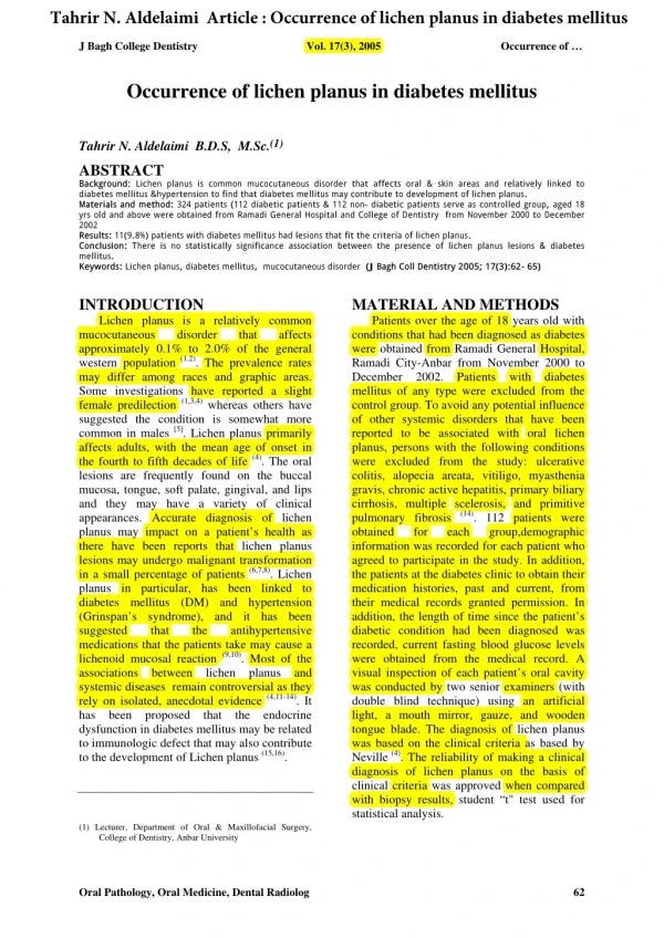
Oral Lichen Planus. Dept. of Oral & Maxillofacial Surgery, Dental Clinic, Gachon Medical School, Ghil Medical Center. Nam Hun Kim. Introduction. 0.5-2.2% 30-60 Years , Female Mild, Chronic, Resolve within 2 years Punctuation, by Acute Exacerbations Oral Neoplasia : Close Observation.

Oral Lichen Planus
E N D
Presentation Transcript
Oral Lichen Planus Dept. of Oral & Maxillofacial Surgery, Dental Clinic, Gachon Medical School, Ghil Medical Center Nam Hun Kim
Introduction • 0.5-2.2% • 30-60 Years, Female • Mild, Chronic, Resolve within 2 years • Punctuation, by Acute Exacerbations • Oral Neoplasia : Close Observation
Etiology • 정확한 원인과 발병기전 밝혀지지 않았음. • 면역 이상의 증거로 상피기저층을 T임파구가 파괴한다는 가설 • 캔디다 감염과 관계있다는 가설 • 신경직적이고 예민한 사람. • C형간염 • 심리적인 고민 • Mercury, gold sodium thiosulphate, palladium chloride 에 대한 감수성 증가.
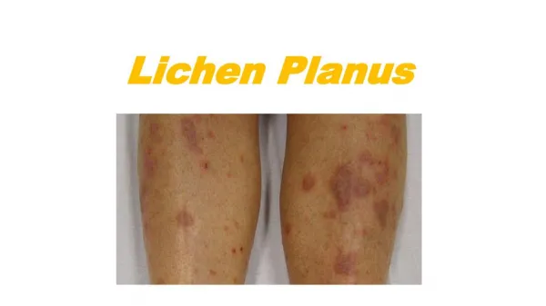
Classification 1)망상형(Reticular type) • 가장 일반적. • 백색구진이 병합되어 몇 개의 가는 하얀 선 또는 작은 구진이 레이스처럼 거미줄 모양으로 됨 (Wickham's striae)
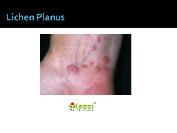
Classification 2)미란형 or 궤양형 (Erosive type) • 두번째로 흔한 형태 • 표면상피의 완전한 상실로 인한 미란의 결과로 발생되며 협점막과 혀에 흔히 나타남 • 소포, 수포( 구진이나 선조의 이면) 가 나타난후 파괴되어 미란이 발생 • 동통성 미란. 치료 필요함.
Classification 3)위축형 (Atrophic type) • 보통 미란형의 결과 • 적색의 비궤양 점막반으로 나타남 • 부착상피 박리성 치은염. (desquamative gingivitis)
Classification 4)플라그형 (Hypertrophic type) • 협점막 , 혀 • 무증상으로 알지 못함. • 고형의 하얀 플라그 • 호발부위 : 협점막
Classification 5) 착색형태 (Pigmented type ) • 극히 드물다. • 백색병소와 망상형태에 배열된 착색된 구진이 특징. • 질환의 급성기 동안 국소적인 멜라닌의 과생산 • 병소치유후 발생할수 있는 착색과 구별.
Diagnosis • 대다수 무증상 • 증상이 있는 경우 치료 (Atrophic, Ulcerative) • 대칭성 • 협점막, 혀, 치은 , 구개, 구강저, 입술
Diagnosis (감별) • Lichenoid lesion, • Leukoplakia, • Lichen sclerosis, • Lupus erythematosus, • chronicUlcerative stomatitis, • Malignant neoplasia
Risk of Malignat Transformation • Controversial • 1.2% • 3 years After Dx. • Current increase state • High risk : 1. Atrophic plaque type 2. Lesion on Tongue, Mouth floor 3. Smoking, Alcoholism
Examination • Histologic biopsy • Immunofluorescence conformation
Management (General Measures) • Oral hygiene: • CHX , antibacterial mouth wash • Dental restoration 에 의한 마찰력 • Past hx : immunosuppressive agent • Saliva collection & culture : Recurrent Candidiasis • Antifungal Tx ( Nystatin Oral suspension Miconasole gel, amphotericin lozenges)
Topical agents • Analgesia : Lidocaine gel, dyphenhydramine • Topical steroids (first of choice) • Triamcinolone : 경미한 증상의 OLP
Topical agents • Fluocinonide : 0.025% • Betamethasone valate : Aerosol type, • Clobetasol proprionate
Topical agents • Topical Cyclosporin 1.Controversial 2. Expensive 3. No commercially preparations
Systemic agent • Prednisolone 1.Topical 로 치료가 안될경우 2.30-60mg 을 2-3 주간 매일 사용
Surgical Techniques • Surgical excision & biopsy • cryotherapy, • CO2 laser, ND:YAG laser
Case Report 1 27Y/F 초진 : Lt. lower buccal area 1개월 전부터 ulceration EX. Bx. 1주일 후 : f/u
Case Report 2 • 37/M • 초진: 4개월 전부터 입안이 쓰리고 아픔 Both lower buccal mucosa ulceration white patch Rx) Oradex . 10A + saline 100cc • 1주일 후: 증상호전
Case Report 3 • 60/F • 초진: 4년전 입안이 벗겨지고 쓰림. white linear striae Mild erythmatous, imflammatory state Rx) Methyl prednisolone po. B-com (Vit. ) • 1주일후 : SX. Retained Rx.) Trental po.
Summary • LP 의 정확한 병인이 밝혀지지 않는 한 Atrophic, erosive lesion 의 증상완화에 목표