Download
1 / 20
220 likes | 314 Vues
Detailed MRI images of the lower thigh, highlighting muscle groups, bone structures, and vascular anatomy. Explore identification of muscles, bones, and nerves in the lower extremity.

E N D
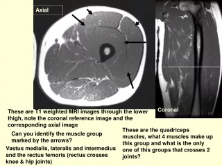
Axial Coronal These are T1 weighted MRI images through the lower thigh, note the coronal reference image and the corresponding axial image These are the quadriceps muscles, what 4 muscles make up this group and what is the only one of this groups that crosses 2 joints? Can you identify the muscle group marked by the arrows? Vastus medialis, lateralis and intermedius and the rectus femoris (rectus crosses knee & hip joints)
What is the structure in the center of the muscle? (dark on outside bright in the center) Cortical bone, black signal, is very densely packed and generates almost no signal on MRI. The marrow space is filled with fat and marrow producing elements and is similar signal to the rest of the fat on MRI images. This is the femur, so why is it dark on the outside and bright on the inside?
Add mag N Hamstrings What is this vascular structure in the medial aspect of the thigh? What nerve lies between the adductor magnus and the hamstrings? Femoral artery What muscle overlies the femoral artery in the thigh? Sciatic nerve Sartorius Muscle
This axial slice is a little lower down. Can you name the vascular structures posterior to the femur? What are the nerves just lateral and posterior to the vessels? These are the popliteal artery and vein. Tibial nerve (solid arrow) Common peroneal nerve (dashed arrow)
Why does the femur have less black (cortex) and more medullary space (marrow fat) down close to the knee joint? This is because we have left the diaphysis and are now in the metaphysis where there is more marrow and cortex is thinner. Look at the cortical thickness
Let’s name some muscles. Can you point out the vastus medialis? Semimembranosis Biceps femoris Semitendinosis, just a tendon at this level Sartorius Gracilis, it is just a tendon here
These are some more inferior images of the knee, other side (left) from the previous patient. Can you follow the popliteal artery and vein and note their relationship to the nerves (tibial and peroneal together at this level). A V N
Can you identify the bone anterior to the femur. This is the patella. Note that the peroneal nerve is starting to head lateral at this level
Note how the muscles change around the knee. The semitendinosis and the gracilis are just tendons (black dots) at this level. Semitendinosis Gracilis
Look at the superficial peroneal nerve at the level. Where is it heading?
Why don’t we see any bones on this image? Because we are at the level of the knee joint
We are now below the level of the knee joint. What bone are we looking at? This is the tibia. Where is the fibula? B
What is this black structure attaching to the tibial tubercle? This is the patellar tendon. Do you see the fibula now?
What are the muscles you see in the upper calf posteriorly? Medial and lateral gastocnemius muscles. This is the solues muscle Popliteus
On this CT angiogram can you identify the popliteal artery, take-off of the anterior tibial artery, peroneal artery and posterior tibial artery? Anterior tibial arteries Peroneal arteries Popliteal arteries Posterior tibial arteries
This is a patient with complete disruption of the distal superficial femoral artery/upper popliteal artery. You can see how it is reconsitiuted (through collaterals) at the level of the knee joint. Disrupted Reconstituted