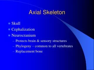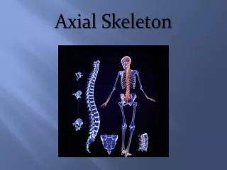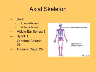
Axial Skeleton
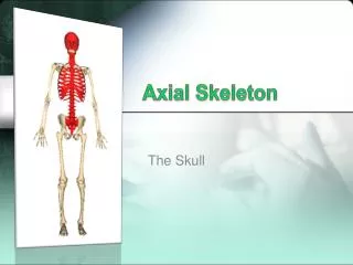
Axial Skeleton. Chapter 6 Part II. The Skull of an Infant. At birth, fusion is ongoing, there are still 2 frontal, 4 occipital, a number of sphenoid, and temporal bone elements - the cranial bones are connected by areas of fibrous CT

Axial Skeleton
E N D
Presentation Transcript
Axial Skeleton Chapter 6 Part II
The Skull of an Infant • At birth, fusion is ongoing, there are still 2 frontal, 4 occipital, a number of sphenoid, and temporal bone elements - the cranial bones are connected by areas of fibrous CT - CT connections are flexible and the skull can be distorted without damage • Fontanels, are the largest fibrous regions between the cranial bones. They include the: - Anterior, Posterior, Sphenoidal, and Mastoid • By age 5 the brain stops growing and the cranial sutures develop
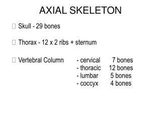
The Vertebral Column • Consists of 26 bones: - 24 vertebra, 1 sacrum, and 1 coccyx • Performs several functions: - provides a column of support - bears the weight of the head, neck, and trunk - transfers weight to the lower limbs - encloses and protects the spinal cord - provides a passageway for spinal nerves - helps maintain the upright position of the body
Regions of the Vertebral Column From superior to inferior, they are • Cervical (7) • Thoracic (12) • Lumbar (5) • Sacral (1) composed of 5 fused vertebrae • Coccygeal (1) composed of 3-5 fused vertebrae
Spinal Curves • Weight-transferring spinal curves are named for the region of the vertebral column they occur in • Primary curves/accommodation curves appear during late fetal development - sacral curve and thoracic curve - allow room for the abdominopelvic viscera • Secondary curves/compensation curves appear several months after birth - lumbar curve and cervical curve - develop as an infant learns to hold up its head and begins to walk • Curves are fully developed by the time a child is 10
Vertebral Column Fig 6.19
Development of Spinal Curves When standing, body weight must be transmitted through the column to the hips and lower limbs. But most of the body weight Lies in front of the column - various curves bring the weight of the body in line with the body axis and its center of gravity Fig 6.19
Vertebral Anatomy Fig 6.21
Cervical Vertebrae • 7 total – smallest, most superior vertebrae • Spinous processes are relatively stumpy and may be split, resulting in a bifid process • Costal processes are extra extensions of bone from the ventrolateral body that attach to the transverse processes • Transverse foamina result from the hole between the costal process and the transverse
Cervical Vertebrae Fig 6.22
C1 - The Atlas • The atlas has no body and articulates cranially with the occipital condyles - these articulations allow one to nod ‘yes’ but prevent twisting • The atlas has 2 arches – the anterior and posterior vertebral arches • Superior and inferior articular facets do not extend beyond the arches
C2 – The Axis • The body of the atlas fuses with the body of the axis during development to form the dens (odontoid process) - because of the dens, there is no intervertebral disc • The articulation between the atlas and axis allows one to shake their head ‘no’
The Axis Fig 6.23
Articulated Atlas and Axis Fig 6.23
C7 - Vertebra Prominens • The last cervical vertebrae, and therefore resembles the thoracic vertebra in structure - has a long, slender spinous process - enlarged transverse processes that may or may not contain a transverse foramen • An elastic ligament called the ligamentum nuchae extends from the spinous process cranially to the occipital crest
Thoracic Vertebrae • 12 total thoracic vertebrae make up the posterior of the rib cage • The bodies have a heart shape • The spinous process is long and slender and points on a posterocaudal angle • The transverse processes point dorsolateral • The thoracic vertebrae articulate with ribs and contain extra facets
Thoracic Vertebrae Fig 6.24
Lumbar Vertebrae • The largest vertebrae (5) make up the lower back region – weight bearing • The body is very thick and oval shaped • Relatively small vertebral foramina are triangular • The transverse processes point more laterally • The spinous process resembles a tail fin of a fish, stumpy and flattened
The Sacrum • Is curved with a convex dorsal surface, consists of the fused components of 5 sacral vertebrae - fuse shortly after puberty (25 – 30) • Protects reproductive, digestive, and excretory organs • Broad surface area provides an extensive area for attachment of muscles - especially those involved in thigh movement • Sacral apex – narrow caudal portion • Broad superior surface forms the base
The Coccyx • Consists of 3 – 5 fused coccygeal vertebrae - begin fusing by age 26 • Provides an attachment site for ligaments and a muscle that constricts the anal opening • Coccygeal cornua – laminae of the 1st vertebra curve to meet the cornua of the sacrum • In males, the adult coccyx points anteriorly while in females it points inferiorly • In the very elderly the coccyx may fuse with the sacrum
The Thoracic Cage Has 2 functions: • Protects the heart, lungs, thymus, and other structures within the cavity • Serves as the attachment site for muscles involved in: - Respiration - Positioning the vertebral column - Movements of the pectoral girdle and upper limb
The Thoracic Cage Fig 6.27
Clinical Terms • Chest tube • Craniostenosis • Deviated nasal septum • Hemothorax • Microcephaly • Pneumothorax • Spina bifida • Whiplash