Axial Skeleton
Lab # 4. Axial Skeleton. Skeleton. Axial Skeleton. Appendicular Skeleton. Axial Skeleton. Skull. Vertebral column. Thoracic cage. Axial Skeleton I. The Skull. 1- Frontal (1) . 2- Parietal (2) . Cranial bones. 3- Temporal (2) . 4- Occipital (1) . 5- Sphenoid (1) .

Axial Skeleton
E N D
Presentation Transcript
Lab # 4 Axial Skeleton
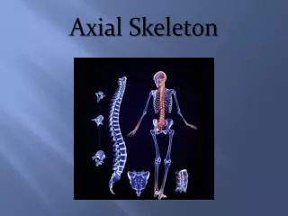
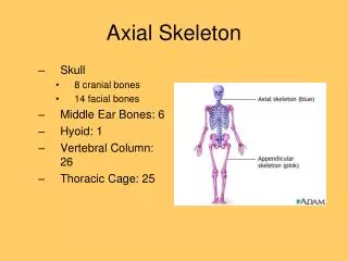
Skeleton Axial Skeleton Appendicular Skeleton
Axial Skeleton Skull Vertebral column Thoracic cage
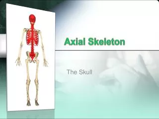
Axial Skeleton I The Skull
1- Frontal (1) 2- Parietal (2) Cranial bones 3- Temporal (2) 4- Occipital (1) 5- Sphenoid (1) 6- Ethmoid (1) 7- Ear ossicles (6) Skull 1- Nasal (2) 2- Lacrimal (2) 3- Inferior nasal concha (2) Facial bones 4- Palatine (2) 5- Vomer (1) 6- Zygomaticus (2) 7- Maxilla (2) 8- Mandible (1)
CRANIAL BONES Coronal suture Squamous suture FRONTAL BONE PARIETAL BONE SPHENOID BONE TEMPORAL BONE ETHMOID BONE NASAL BONE OCCIPITAL BONE LACRIMAL BONE External auditory canal ZYGOMATIC BONE Mandibular fossa MAXILLA Mastoid process Styloid process Condyloid process of mandible MANDIBLE Lateral view
The Skull Coronal suture Squamous suture FRONTAL BONE PARIETAL BONE SPHENOID BONE TEMPORAL BONE ETHMOID BONE NASAL BONE Mandibular fossa External auditory canal LACRIMAL BONE OCCIPITAL BONE ZYGOMATIC BONE MAXILLA Alveolar process of maxilla Mastoid process Styloid process Condyloid process of mandible Alveolar process of mandible MANDIBLE CRANIAL BONES FACIAL BONES Lateral view
Nasal septum Vomer Perpendicular plate of ethmoid Middle nasal concha Alveolar process of maxilla Inferior nasal concha Alveolar process of mandible
FRONTAL BONE NASAL BONE PARIETAL BONE TEMPORAL BONE SPHENOID BONE LACRIMAL BONE ETHMOID BONE ZYGOMATIC BONE Perpendicular plate of ethmoid bone INFERIOR NASAL CONCHA VOMER Alveolar processes of maxilla MAXILLA Anterior view Middle nasal concha (part of ethmoid) Alveolar processes of mandible MANDIBLE CRANIAL BONES FACIAL BONES
Hypophyseal fossa or sellaturcica SPHENOID CRANIAL BONES Cribriform plate of ethmoid bone Cristagalli of ethmoid bone Carotid canals Jugular foramen
Palatine process of maxilla Cribriform plate of ethmoid bone Hard palate Crista galli of ethmoid bone PALATINE BONE Hypophyseal fossa Anterior cranial fossa Optic foramina SPHENOID BONE SPHENOID BONE Foramen ovale Foramen lacerum VOMER BONE Middle cranial fossa Foramen rotundum Foramen ovale Carotid canal Foramen spinosum TEMPORAL BONE Mandibular fossa Foramen lacerum Styloid process Occipital condyles Mastoid process Posterior cranial fossa OCCIPITAL BONE OCCIPITAL BONE Foramen magnum Inferior view Interior view Jugular foramen
CRANIAL AND FACIAL BONES ARE CONNECTED BY JOINTS, WHICH ARE INMOVABLE AND ARE REFERRED AS SUTURES 1- Sagittal suture 2- Coronal suture 3- Lambdoid suture 4- Squamous suture
SUTURES Sagittal suture Lambdoid suture Coronal suture PARIETAL BONE (right) PARIETAL BONE (left) OCCIPITAL BONE FRONTAL BONE Posterior view Superior view
SUTURES Squamous suture PARIETAL BONE FRONTAL BONE SPHENOID TEMPORAL BONE OCCIPITAL BONE ETHMOID
FRONTAL BONE Coronal suture Sagittal suture PARIETAL BONE PARIETAL BONE Lambdoid suture PARIETAL BONE PARIETAL BONE OCCIPITAL BONE TEMPORAL BONE MANDIBLE Mastoid process Lambdoid suture Posterior view Superior view OCCIPITAL BONE
SINUSES: They are air-filled chambers in the interior of some bones Sphenoidal sinus Hypophyseal fossa PARIETAL Cristagalli FRONTAL TEMPORAL OCCIPITAL SPHENOID ETHMOID Frontal sinus VOMER Cribriform plate of ethmoid MAXILLA Perpendicular plate of ethmoid Mastoid process Styloid process PALATINE BONE Palatine process of maxilla Midsagittal view
PARIETAL Hypophyseal fossa FRONTAL Sphenoidal sinus Crista galli Frontal sinus TEMPORAL ETHMOID Ethmoidal sinus SPHENOID Cribriform plate of ethmoid OCCIPITAL Perpendicular plate of ethmoid Palatine process of maxilla Styloid process PALATINE BONE Mastoid process Midsagittal view
The Mandible Mandibular condyle Mandibular notch Coronoid process Mandibular foramen Ramus of mandible Alveolar margin Mental foramen Lateral view Mandibular angle
Posterolateral or mastoid fontanel Posterior or occipital fontanel Anterolateral or sphenoid fontanel Anterior or frontal fontanel FONTANELS: Are areas of fibrous tissue that connect the cranial bones of the infant. The connection are quite flexible, so the skull can be distorted during delivery without damage
Fetal Skull Anterior fontanel Squamous suture Coronal suture Parietal bone Frontal bone Posterior fontanel Anterolateral (sphenoid) fontanel Sphenoid bone Lambdoidal suture Temporal bone Occipital bone Posterolateral (mastoid) fontanel Lateral View of Fetal Skull
Spinous process Vertebral foramen Transverse process Vertebra (prototype) Superior articular surface Inferior articular surface Vertebral body Inferior view
Dens of axis Transverse ligament Atlas Axis Superior articular facet
Vertebral Column of an Adult Cervical curve C7 T1 Atlas (C1) Thoracic curve Axis (C2) Intervertebral discs T12 L1 Lumbar curve L5 Sacrum Pelvic curve Intervertebral foramina Coccyx Lateral view
Atlas (C1) Axis (C2) Spinous process Posterior arch Transverse process Transverse process Lamina Superior articular facet Vertebral foramen Vertebral foramen Transverse foramen Superior articular facet Dens Anterior arch Superior view Superior view
Cervical Vertebra Bifid tip of spinous process Superior articular process Superior articular process Spinous process Superior articular facet Vertebral foramen Body Body Inferior articular process Spinous process Transverse process Transverse process Transverse foramen Superior view Lateral view
Thoracic Vertebra Spinous process Superior articular process Transverse costal facet for tubercle of rib Superior costal demifacet for head of rib Superior articular facet Vertebral foramen Body Superior costal demifacet for head of rib Transverse process Inferior costal demifacet for head of rib Body Inferior notch Spinous process Superior view Lateral view
Lumbar Vertebra Superior articular process Spinous process Spinous process Superior articular process Superior articular facet Vertebral foramen Body Transverse process Body Inferior articular process Superior view Lateral view
A comparison of vertebrae from cervical, thoracic and lumbar regions Superior view Lateral view Cervical (7) Thoracic (12) Lumbar (5)
Transverse foramina of cervical vertebrae are passageways for vertebral artery (and vein, no shown) Transverse foramina Intervertebral foramina Intervertebral foramina are passageways for spinal nerves running to or from the enclosed spinal cord Together, the vertebral foramina of successive vertebrae form the vertebral canal , which encloses the spinal cord Vertebral foramen
Sacrum and Coccyx Superior articular processes Sacrum (Fused components of 5 vertebrae) S1 Median sacral crest Lateral sacral crest S2 S3 S4 Anterior view Posterior view Sacral foramina S5 Sacral hiatus Coccyx (3 to 5 coccygeal vertebrae) Coccyx
The Thoracic Cage T1 1 1 1 1 2 2 2 2 Costal cartilages 3 3 3 3 True or vertebrosternal ribs (ribs 1-7) 4 4 4 4 They are connected to the sternum by separated costal cartilages 5 5 5 5 6 6 6 6 7 7 7 7 T12 8 8 Vertebrochondral (8,9,10) False ribs (ribs 8-12) 9 9 L1 Floating or vertebral (11,12) 10 10 11 12 12 11 Floating ribs Floating ribs
Sternum Rib Tubercle Anterior (sternal) end Manubrium Posterior (vertebral) end Neck Head Articulation between the ribs and thoracic vertebrae Body Transverse costal facet Tubercle Neck Head Demifacet Xiphoid process Anterior view Superior view
Sternum Sternum
Movements of curve ribs affects the volume of the thoracic cage Inhalation Rib cage elevates and diaphragm contracts Exhalation Rib cage returns to the normal position and diaphragm relaxes