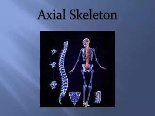
Axial Skeleton
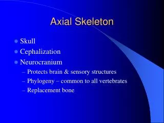
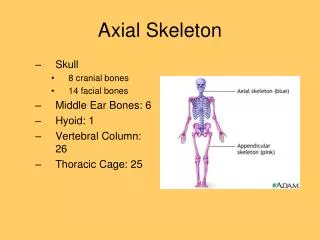
Axial Skeleton. Skull. Cranial Bones Facial Bones. Cranial Bones. Frontal Bone – Forms the forehead, the roofs of the orbits, and most of the anterior part of the cranial floor. Cranial Bones. Parietal Bones – form the greater portion of the sides and roof of the cranial cavity.

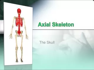
Axial Skeleton
E N D
Presentation Transcript
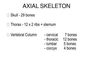
Skull • Cranial Bones • Facial Bones
Cranial Bones • Frontal Bone – Forms the forehead, the roofs of the orbits, and most of the anterior part of the cranial floor
Cranial Bones • Parietal Bones – form the greater portion of the sides and roof of the cranial cavity
Cranial Bones • Temporal Bones – Form the inferior lateral aspects of the cranium and part of the cranial floor
Cranial Bones • Occipital Bone – Forms the posterior part and most of the base of the cranium
Cranial Bones • Sphenoid Bone – Lies at the middle part of the base of the skull
Cranial Bones • Ethmoid Bone – a light, sponge-like bone located on the midline in the anterior part of the cranial floor medial to the orbits.
Facial Bones • Nasal Bones – meet at the midline and form part of the bridge of the nose
Facial Bones • Maxillae – unit to form the upper jaw.
Cleft Palate • If the maxillary bones do not unite during the weeks 10 to 12 of embryonic development, cleft palate occurs.
Facial Bones • Zygomatic Bones – (cheekbones) form the prominence of the cheeks and part of the lateral wall and floor of each orbit.
Facial Bones • Lacrimal Bones – They are posterior and lateral to the nasal bones and form part of the medial wall of each orbit.
Facial Bones • Palatine Bones – They form the posterior part of the hard palate, part of the floor and lateral wall of the nasal cavity, and a small part of the floor of the orbits.
Facial Bones • Inferior Nasal Concha – form a portion of the inferior lateral wall of the nasal cavity and project into the nasal cavity
Facial Bones • Vomer – A rough triangular bone on the floor of the nasal cavity
Facial Bones • Mandible – lower jawbone
Nasal Septum • Divides the nasal cavity into R. and L. sides • Consists of vomer, septal cartilage (hyaline cartilage), and perpendicular plate of the ethmoid bone
Deviated nasal septum • This occurs when the nasal septum is deflected laterally from the midline of the nose
Orbits Made up of seven bones; • Frontal • Sphenoid • Ethmoid • Palatine • Zygomatic • Lacrimal • Maxilla
Paranasal Sinus • Paired cavities found in the frontal, sphenoid, ethmoid, and maxillary
Paranasal Sinus • Lined with mucous membranes that are continuous with the lining of the nasal cavity
Fontanels • Soft spots – membrane filled spaces
Fontanels • Allow rapid growth of brain during infancy
Fontanels • Allow skull to change shape as it passes through the birth canal
Fontanels • Bone ossification occurs via intramembranous
Hyoid Bone • Between mandible and larynx
Hyoid Bone • Not adam’s apple
Hyoid Bone • Suspended from the styloid processes of the temporal bones by ligaments and muscles.