Cell structure and function Chapter 3
Cell structure and function Chapter 3. Processes of Life. Growth Reproduction Responsiveness Metabolism. Prokaryotes. Do not have membrane surrounding their DNA; no nucleus Lack various internal structures bound with phospholipid membranes Small; ~1.0 µm in diameter Simple structure

Cell structure and function Chapter 3
E N D
Presentation Transcript
Processes of Life • Growth • Reproduction • Responsiveness • Metabolism
Prokaryotes • Do not have membrane surrounding their DNA; no nucleus • Lack various internal structures bound with phospholipid membranes • Small; ~1.0 µm in diameter • Simple structure • Comprised of bacteria and archaea
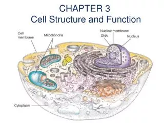
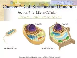
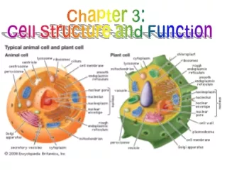
Eukaryotes • Have membrane surrounding DNA; have nucleus • Have internal membrane-bound organelles • Are larger; 10-100 µm in diameter • Have more complex structure • Comprised of algae, protozoa, fungi, animals, and plants
Comparing Prokaryotes and Eukaryotes Figure 3.2a
Comparing Prokaryotes and Eukaryotes Figure 3.2b
External Structures of Prokaryotic Cells • Glycocalyces • Flagella • Fimbriae and pili
Glycocalyces • Gelatinous, sticky substance surrounding the outside of the cell • Composed of polysaccharides, polypeptides, or both • Two types • Capsule • Slime layer
Capsule • Composed of organized repeating units of organic chemicals • Firmly attached to cell surface • Protects cells from drying out • May prevent bacteria from being recognized and destroyed by host
Example of Capsule Figure 3.4a
Slime Layer • Loosely attached to cell surface • Water soluble • Protects cells from drying out • Sticky layer that allows prokaryotes to attach to surfaces
Example of Slime Layer Figure 3.4b
Flagella • Are responsible for movement • Have long structures that extend beyond cell surface • Not all prokaryotes have flagella
Bacterial Flagella Structure • Composed of filament, hook, and basal body • Flagellin protein (filament) is deposited in a helix at the lengthening tip • Base of filament inserts into hook • Basal body anchors filament and hook to cell wall by a rod and a series of either two or four rings of integral proteins • Filament capable of rotating 360º
Bacterial Flagella Structure Figure 3.5a
Bacterial Flagella Structure Figure 3.5b
Arrangements of Bacterial Flagella Figure 3.6a
Arrangements of Bacterial Flagella Figure 3.6b
Arrangements of Bacterial Flagella Figure 3.6c
Function of Bacterial Flagella • Rotation propels bacterium through environment • Rotation can be clockwise or counterclockwise; reversible • Bacteria move in response to stimuli (taxis) • Runs – movements of cell in single direction for some time due to counterclockwise flagellar rotation; increase with favorable stimuli (positive chemotaxis, positive phototaxis) • Tumbles – abrupt, random, changes in direction due to clockwise flagellar rotation; increase with unfavorable stimuli (negative chemotaxis, negative phototaxis)
Fimbriae and Pili • Nonmotile extensions • Fimbriae • Sticky, proteinaceous, bristlelike projections • Used by bacteria to adhere to one another, to hosts, and to substances in environment • May be hundreds per cell and are shorter than flagella • Serve an important function in biofilms
Fimbriae Versus Flagella Figure 3.9
Pili • Long hollow tubules composed of pilin • Longer than fimbriae but shorter than flagella • Bacteria typically only have one or two per cell • Join two bacterial cells and mediate the transfer of DNA from one cell to another (conjugation) • Also known as conjugation pili or sex pili
Pilus Versus Fimbriae Figure 3.10
Prokaryotic Cell Wall • Provides structure and shape and protects cell from osmotic forces • Assists some cells in attaching to other cells or in eluding antimicrobial drugs • Animal cells do not have; can target cell wall of bacteria with antibiotics • Bacteria and archaea have different cell wall chemistry
Bacterial Cell Wall • Most have cell wall composed of peptidoglycan; a few lack a cell wall entirely • Peptidoglycan composed of sugars, NAG, and NAM • Chains of NAG and NAM attached to other chains by tetrapeptide crossbridges • Bridges may be covalently bonded to one another • Bridges may be held together by short connecting chains of amino acids • Scientists describe two basic types of bacterial cell walls: gram-positive and gram-negative
Gram-Positive Cell Wall • Relatively thick layer of peptidoglycan • Contains unique polyalcohols called teichoic acids • Some covalently linked to lipids, forming lipoteichoic acids that anchor peptidoglycan to cell membrane • Retains crystal violet dye in Gram staining procedure; appear purple • Acid-fast bacteria contain up to 60% mycolic acid; helps cells survive desiccation
Gram-Negative Cell Walls • Have only a thin layer of peptidoglycan • Bilayer membrane outside the peptidoglycan contains phospholipids, proteins, and lipopolysaccharide (LPS) • May be impediment to the treatment of disease • Following Gram staining procedure, cells appear pink
LPS • Union of lipid with sugar • Also known as endotoxin • Lipid portion known as lipid A • Dead cells release lipid A when cell wall disintegrates • May trigger fever, vasodilation, inflammation, shock, and blood clotting • Can be released when antimicrobial drugs kill bacteria
Periplasmic Space • Located between outer membrane and cell membrane • Contains peptidoglycan and periplasm • Contains water, nutrients, and substances secreted by the cell, such as digestive enzymes and proteins involved in transport
Bacterial Cell Walls Figure 3.13a
Thick layer peptidoglycan • Teichoic acid • Lipoteichoic acid Gram Positive Cell Wall
Bacterial Cell Walls Figure 3.13b
Outer Membrane • LPS-lipopolysaccharide (Endotoxin) • Porins • Periplasm Gram Negative Cell wall
Archael Cell Walls • Do not have peptidoglycan • Cell walls contain variety of specialized polysaccharides and proteins • Gram-positive archaea stain purple • Gram-negative archaea stain pink
Prokaryotic Cytoplasmic Membrane • Referred to as phospholipid bilayer; composed of lipids and associated proteins • Approximately half the membrane is composed of proteins that act as recognition proteins, enzymes, receptors, carriers, or channels • Integral proteins • Peripheral proteins • Glycoproteins • Fluid mosaic model describes current understanding of membrane structure
Phospholipid Bilayer of Cytoplasmic Membrane Figure 3.14
Cytoplasmic Membrane Function • Controls passage of substances into and out of the cell; selectively permeable • Harvests light energy in photosynthetic prokaryotes
Control of Substances Across Cytoplasmic Membrane • Naturally impermeable to most substances • Proteins allow substances to cross membrane • Occurs by passive or active processes • Maintains a concentration gradient and electrical gradient • Chemicals concentrated on one side of the membrane or the other • Voltage exists across the membrane
Passive Processes of Transport • Diffusion • Facilitated diffusion • Osmosis • Isotonic solution • Hypertonic solution • Hypotonic solution
Effects of Solutions on Organisms Figure 3.18
Active Processes of Transport • Active Transport • Utilizes permease proteins and expends ATP • Uniport • Antiport • Symport • Group Translocation • Substance chemically modified during transport
Cytoplasm of Prokaryotes • Cytosol – liquid portion of cytoplasm • Inclusions – may include reserve deposits of chemicals • Ribosomes – sites of protein synthesis • Cytoskeleton – plays a role in forming the cell’s basic shape • Some bacterial cells produce dormant form called endospore
Endospores • Hardiest of all life forms • For escape from unfavorable environmental conditions • Germination = return to the vegetative state from the spore state • NOT reproductive (1 cell forms 1 endospore which return to reform 1 cell)
External Structure of Eukaryotic Cells • Glycocalyces • Never as organized as prokaryotic capsules • Helps anchor animal cells to each other • Strengthens cell surface • Provides protection against dehydration • Function in cell-to-cell recognition and communication
Eukaryotic Cell Walls • Fungi, algae, plants, and some protozoa have cell walls but no glycocalyx • Composed of various polysaccharides • Cellulose found in plant cell walls • Fungal cell walls composed of cellulose, chitin, and/or glucomannan • Algal cell walls composed of cellulose, proteins, agar, carrageenan, silicates, algin, calcium carbonate, or a combination of these • …..NO peptidoglycan
Eukaryotic Cytoplasmic Membrane • All eukaryotic cells have cytoplasmic membrane • Is a fluid mosaic of phospholipids and proteins • Contains steroid lipids to help maintain fluidity • Controls movement into and out of cell