Download
1 / 30
300 likes | 499 Vues
Learn about the differentiation of the ovary from primary sex cords to secondary sex cords. Understand the development of the ovary and paramesonephric ducts leading to the formation of the female genital tract.

E N D
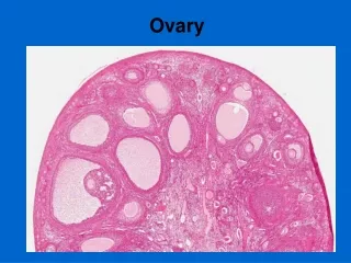
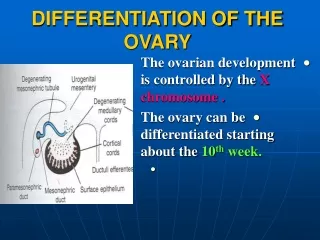
DIFFERENTIATION OF THE OVARY • The ovarian development is controlled by the X chromosome . • The ovary can be differentiated starting about the 10th week.
PRIMARY SEX CORDS • They dissociate into irregular cell clusters • Which extend into the medulla where they form the rete ovarii. • Normally, the cords and the rete ovarii degenerate and disappear.
SECONDARY (CORTICAL) SEX CORDS • They are formed from the proliferating surface epithelium. • They penetrate the mesenchyme but they still close to the surface epithelium.
SECONDARY SEX CORDS • In the 4th month (16weeks) the secondary sex cords split into isolated cell clusters (primordial follicles).
PRIMORDIAL FOLLICLES • Each follicle contains oogonium ( from the primitive germ cells ) It is surrounded by a single layer of flattened cells (follicular cells) derived from the sex cords. • Active mitosis of oogonia produces thousands of the primordial follicles.
PRIMORDIAL FOLLICLES • No oogonia form postnatally. • Before Birth: • The oogonia enlarge to form the primary oocytes.
PRIMORDIAL FOLLICLES • After birth ,the surface epithelium becomes flattened into a single layer. • It is separated from the follicles in the cortex by a thintunica albuginea.
DESCENT OF THE OVARIES • The ovaries descend from the posterior abdominal wall into the pelvis. • The Gubernaculum is attached to the uterus near the attachment of the uterine tubes.
DESCENT OF THE OVARIES • The cranial part of the Gubernaculum becomes the ovarian ligament. • The caudal part becomes the round ligament of the uterus. • The round ligament passes in the inguinal canal and terminates in the labia majora.
PARAMESONEPHRIC DUCTS • They form most of the female genital tract. • They develop because of the absence of MIS. • They are formed from longitudinal invagination of epithelium lateral to the mesonephroi.
PARAMESONEPHRIC DUCTS • Cranially: • Their funnel shaped ends open into the peritoneal cavity. • Caudally : • They run lateral to the mesonephric ducts. • They Cross them ventrally. They descend caudo medially and approach each other in the median plane. • They form the y- shaped uterovaginal primordium.
UTEROVAGINAL PRIMORDIUM • It opens in the dorsal wall of the urogenital sinus and produces sinus (mullerian)tubercle.
DERIVATIVES OF PARAMESONEPHRIC DUCTS • 1. UTERINE TUBES • They develop from the cranial un fused parts of the paramesonephric ducts.
DERIVATIVES OF PARAMESONEPHRIC DUCTS • 2. UTEROVAGINAL PRIMORDIUM • It differentiates into • uterus (body and cervix) and superior part of the vagina.
PARAMESONEPHRIC REMNANTS IN (MALES) • Its cranial end may persist as the appendix of the testis (attached to the superior pole of the testis). • The prostatic utricle is homologus to thevagina.
PERITONEUM • Movement of the paramesonephric ducts mediocaudally shifts the urogenital ridges to lie in a transverse plane. • Fusion of the caudal ends of the ducts brings • (1) Two peritoneal pouches: • Rectouterine. Vesicouterine.
PERITONEUM • (2) Two peritoneal folds: Right and Left Broad ligaments.
STRUCTURE OF UTERUS • The endometrial stroma and myometrium of the uterus are derived from the adjacent splanchnic mesenchyme. • The mesenchyme differentiate between the layers of the broad ligament to form the parametrium.
VAGINA • The vagina has a dual origin : • (1) upper portion from the uterovaginal priordium. • (2) lower portion from the urogenitalsinus.
VAGINA • The solid tip of the paramesonephric ducts at the urogenital sinus (sinus tubercle) produce two solid evaginations (SinoVaginal Bulbs) they grow out from the pelvic part of the sinus.
VAGIN (LOWER PART) • The bulbs will proliferate and fuse to form a solid Vaginal Plate. • The Proliferation continues and it increases the distance between the urogenital sinus and the uterus.
VAGINAL PLATE • The central cells of the vaginal plate break down to form the lumen of the vagina. • The lining of the entire vagina is from the vaginal plate.
VAGINAL FORNICES • They are the wing like expansions of the vagina around the end of the uterus. • They are of paramesonephric origin.
HYMEN • It is a thin plate of tissue that separates the lumen of the vagina from that of the urogenital sinus. • It is formed from invagination of the posterior wall of the urogenital sinus due to expansion of the caudal end of the vagina.
HYMEN • It usually ruptures during the perinatal period. • It remains as a thin fold of mucous membrane just within the vaginal orifice.
URETHRAL & PARAURETHRAL GLANDS • They grow from the urethra into the surrounding mesenchyme. • They are corresponding to the prostate gland of the male.
GREATER VESTIBULAR (BARTHOLIN) • They are outgrowths of the urogenital sinus. • They are corresponding to the bulbourethral glands of the male.
REMNANTS OF MESONEPHRIC DUCTS • They degenerate because of lack of testosterone. • They are represented by: • 1. Epoophoron : Efferent ductules and duct of epididymis in male. • 2. Paroophoron : Rudimentary tubules closer to the uterus.
REMNANTS OF MESONEPHRIC DUCTS • 3. Duct of Gartner : • It lies between the layers of the broad ligament. • It correspondes to the ductus deferens and ejaculatory duct of male.