
Pearl Analysis and Differentiation Techniques: A Comprehensive Guide
E N D
Presentation Transcript
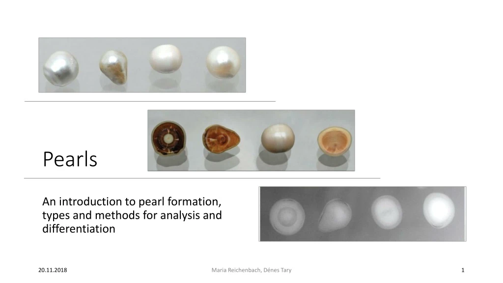
Pearls An introduction to pearl formation, types and methods for analysis and differentiation Maria Reichenbach, DénesTary
Overview Maria Reichenbach, DénesTary – Pearls • Introduction to pearls • "Household methods" • Methods of visualization • X-ray radiography (2D) • X-ray tomography (3D) • Neutron imaging • X-ray luminescence – salt- vs. freshwater pearls • Origin of a pearl • DNA analysis • Fluorescence • Age of pearl: Radiocarbon analysis
The givenproblem https://i2.wp.com/deleusejewelers.com/wp-content/uploads/2010/08/IMG_00011.jpg?fit=492%2C360 Maria Reichenbach, DénesTary – Pearls 3 Pearl pendant as heritage Not mentioned in will Value?
Whatis a pearl? Hänni, H.A., 2012. Natural pearls and cultured pearls: a basic concept and its variations. The Australian Gemmologist, 24(11), pp.256-266. Maria Reichenbach, DénesTary – Pearls 4 Biological process in bivalves Accidental Reaction against irritant (tiny organism, organic substance)
Typesofpearls • Cultured • Nacre • Withorwithoutbead • Imitated • Plastic, glass https://sc02.alicdn.com/kf/HTB1CLemJVXXXXX9aXXXq6xXFXXXN/NATURAL-PEARLS-OF-PINCTADA-MAXIMA-CULTURED-PEARLS.jpg_350x350.jpg https://i.etsystatic.com/9797693/d/il/987da8/1299888772/il_340x270.1299888772_hd26.jpg?version=0 https://5.imimg.com/data5/LV/NT/MY-38792203/plastic-white-pearl-500x500.jpg Maria Reichenbach, DénesTary – Pearls 5 • Natural • Nacre • Withoutbead
"Housholdmethods" forpearlanalysis • Texture • rub pearl against your teeth or each other. Nacre is sandy, imitates are smooth • Shape • imitates are perfectly spherical, natural pearls are not • Weight • Imitates are not made of nacre so they have the wrong density Maria Reichenbach, DénesTary – Pearls 6 • Temperature • nacre is cold to the touch and warms up with skin contact, but imitates have room temperature
X-Ray radiography Collimator Scintillator Optional filter(s) X-ray source Detector (CCD) Sample Hänni, H.A., 2012. Natural pearls and cultured pearls: a basic concept and its variations. The Australian Gemmologist, 24(11), pp.256-266. Maria Reichenbach, DénesTary – Pearls 7 Project a picture (2D) of the inside structure
X-Ray radiography Meyer, J.B., Cartier, L.E., Pinto-Figueroa, E.A., Krzemnicki, M.S., Hänni, H.A. and McDonald, B.A., 2013. DNA fingerprinting of pearls to determine their origins. PloS one, 8(10), p.e75606 Maria Reichenbach, DénesTary – Pearls 8 • Revelation ofgrowthstructures • Known, individual development • Bead? cultured • Nobead and freshwaterusuallycultured
X-Ray radiography Krzemnicki, M.S., Friess, S.D., Chalus, P., Hänni, H.A. and Karampelas, S., 2010. X-Ray Computed Microtomography: Distinguishing Natural Pearls from Beaded and Non-Beaded Cultured Pearls. Gems & Gemology, 46(2), 128-134. Hänni, H.A., 2012. Natural pearls and cultured pearls: a basic concept and its variations. The Australian Gemmologist, 24(11), pp.256-266. Maria Reichenbach, DénesTary – Pearls 9
X-Ray tomography Collimator Scintillator Optional filter(s) Computer X-ray source Detector (CCD) Sample Maria Reichenbach, DénesTary – Pearls 10 Almostidenticalto x-rayradiography, but 3D More detailwithmicrotomography Betterviewanglesaccessible
X-Ray tomography Krzemnicki, M.S., Friess, S.D., Chalus, P., Hänni, H.A. and Karampelas, S., 2010. X-Ray Computed Microtomography: Distinguishing Natural Pearls from Beaded and Non-Beaded Cultured Pearls. Gems & Gemology, 46(2), 128-134. Maria Reichenbach, DénesTary – Pearls 11 Identifying a non-beadedculturedpearl
Neutron imaging Neutron tomography X-raymicro-tomography Mannes, D., Hanser, C., Krzemnicki, M., Harti, R.P., Jerjen, I. and Lehmann, E., 2017. Gemmological investigations on pearls and emeralds using neutron imaging. Physics Procedia, 88, pp.134-139. Maria Reichenbach, DénesTary – Pearls 12 Expensive, rarelyused Results with comparable image quality Different contrast, highlighting regions containing organic material (hydrogen containing) Such regions are hard to distinguish from voids using X-ray tomography complementary measurement
Salt- orfreshwaterpearls?X-rayluminescence Collimator Optional filter(s) X-ray source Sample Maria Reichenbach, DénesTary – Pearls 13 Luminescence is the spontaneous emission of light not caused by heat Apparatus Conventional X-raycabinet Lead glasswindowtoobservetheluminescence
Salt- or freshwater pearls?X-ray luminescence Hänni, H.A., Kiefert, L., Giese, P., 2005. X-ray luminescence, a useful test in pearl testing Maria Reichenbach, DénesTary – Pearls 14 Fresh-water pearls produce luminescence under X-rays Salt-water pearls do not Fresh-water nacre contains traces of manganese
Origin of a pearl: DNA sequencing • 92% CaCO3: aragonite, calcite, vaterite • 4% organic matter: conchiolin and porphirins contains different types of proteins, makes the colour • 4% water • DNA in organic part between layers • Works for coloured and "white" pearls Negatively charged DNA molecules have high affinity for the Ca2+- ions of CaCO3, which might enhance the conservation of DNA in biogenic gems such as pearls. Meyer, J.B., Cartier, L.E., Pinto-Figueroa, E.A., Krzemnicki, M.S., Hänni, H.A. and McDonald, B.A., 2013. DNA fingerprinting of pearls to determine their origins. PloS one, 8(10), p.e75606 Maria Reichenbach, DénesTary – Pearls 15
Origin of a pearl: DNA sequencing sample preparation • Minimally invasive (10-20 mg) Iftherealreadyis a hole • Powderissuspended in 1000-2000 µL EDTA (0.5 M at pH 8) • Vortexing (thoroughmixing) • Incubateovernight in waterbath at 56°C Dissolvingof CaCO3requiresharshconditions Meyer, J.B., Cartier, L.E., Pinto-Figueroa, E.A., Krzemnicki, M.S., Hänni, H.A. and McDonald, B.A., 2013. DNA fingerprinting of pearls to determine their origins. PloS one, 8(10), p.e75606 Maria Reichenbach, DénesTary – Pearls 16
Origin of a pearl: DNA sequencing:RFLP - restriction fragment length polymorphismus Meyer, J.B., Cartier, L.E., Pinto-Figueroa, E.A., Krzemnicki, M.S., Hänni, H.A. and McDonald, B.A., 2013. DNA fingerprinting of pearls to determine their origins. PloS one, 8(10), p.e75606 Maria Reichenbach, DénesTary – Pearls 17 DNA is cut by restriction enzymes Fragments are ordered by length using gel-electrophoresis DNA samples are compared with each other
Origin of a pearl: provenance from Arabian Gulf? http://species-identification.org/species.php?species_group=caribbean_diving_guide&id=417 Maria Reichenbach, DénesTary – Pearls 18 Pinctada radiata is typical for Arabian Gulf Typically oval pearl shape Compare gained DNA with reference
Origin of a pearl: Laser induced fluorescence – apparatus MyeongJin Ju et al: Multimodal analysis of pearls and pearl treatments by using optical coherence tomography and fluorescence spectroscopy ; OSA, 2011, Vol. 19, No. 7 Maria Reichenbach, DénesTary – Pearls 19
Origin of a pearl: Laser induced fluorescence – used pearls MyeongJin Ju et al: Multimodal analysis of pearls and pearl treatments by using optical coherence tomography and fluorescence spectroscopy ; OSA, 2011, Vol. 19, No. 7 Maria Reichenbach, DénesTary – Pearls 20
Origin of a pearl: Laser induced fluorescence – Time integrated fluorescence spectra MyeongJin Ju et al: Multimodal analysis of pearls and pearl treatments by using optical coherence tomography and fluorescence spectroscopy ; OSA, 2011, Vol. 19, No. 7 Maria Reichenbach, DénesTary – Pearls 21 Akoya South Sea Tahitian Fresh-water (527 nm) (548 nm) (620, 652 nm) (602 nm)
Origin of a pearl: Laser induced fluorescence –Time integrated fluorescence spectra MyeongJin Ju et al: Multimodal analysis of pearls and pearl treatments by using optical coherence tomography and fluorescence spectroscopy ; OSA, 2011, Vol. 19, No. 7 Maria Reichenbach, DénesTary – Pearls 22 South Sea Black Tahitian White Tahitian • Colour ofwhite Tahitian and south sea can not be distinguished by eye but with fluorescence
Origin of a pearl: Laser induced fluorescence –Time integrated fluorescence spectra MyeongJin Ju et al: Multimodal analysis of pearls and pearl treatments by using optical coherence tomography and fluorescence spectroscopy ; OSA, 2011, Vol. 19, No. 7 Maria Reichenbach, DénesTary – Pearls 23 Wavelengths do not overlap with each other Nacre property of a pearl highly depends on the place where the mother oyster was grown
Origin of a pearl: Laser induced fluorescence – decay rates MyeongJin Ju et al: Multimodal analysis of pearls and pearl treatments by using optical coherence tomography and fluorescence spectroscopy ; OSA, 2011, Vol. 19, No. 7 Maria Reichenbach, DénesTary – Pearls 24 Measure for 60 s with 1 s inbetween Cooler conditions cause Akoya pearls to develop their nacre layer more slowly but with more compact crystal structure Affects optical properties and the decay rate of fluorescence
Age of a pearl Krzemnicki, M.S. and Hajdas, I., 2013. Age determination of pearls: A new approach for pearl testing and identification. Radiocarbon, 55(3), pp.1801-1809 Maria Reichenbach, DénesTary – Pearls 25 • Radio carbon dating • Issues: • Sample invasive(?) • Global 14C distribution • System not always closed • Good method for known pearls, difficult otherwise
Summary & conclusions determinationofmothermollusk origin Maria Reichenbach, DénesTary – Pearls 26 • Methods foranalysis • Visualizationgrowthstructure origin • X-rayradiography/(micro)tomography • Neutron tomography • X-rayluminescencesalt- orfreshwaterdifferentiation • Laser inducedfluorescence • DNA sequencing • Radiocarbondatingagehistoricalconclusions • Trainedeye & knowledgeofpearlsisneeded • Growingdifficultiesforthefield due to well-craftedbeadlesspearls • Impossible totellculturedfromnatural in thefuturevisually? Laser inducedfluorescence, DNA sequencing & radiocarbondatingwinrelevance
Questions Maria Reichenbach, DénesTary – Pearls
Gemstones Maria Reichenbach, DénesTary – Pearls 28 • What are the differences between testing a coloured gemstone from pearls? • Translucency allows spectroscopic methods • (Crystal) structure can be analysed • Element compositions vary: elemental analysis • different problem, different strategies • One mentioned parallel strategy: • Neutron tomography to identify enclosed organic matters
Reference List Maria Reichenbach, Dénes Tary – Pearls Hänni, H.A., Kiefert, L., Giese, P., 2005. X-ray luminescence, a useful test in pearl testing Hänni, H.A., 2012. Natural pearls and cultured pearls: a basic concept and its variations. The Australian Gemmologist, 24(11), pp.256-266. Krzemnicki, M.S., Friess, S.D., Chalus, P., Hänni, H.A. and Karampelas, S., 2010. X-Ray Computed Microtomography: Distinguishing Natural Pearls from Beaded and Non-Beaded Cultured Pearls. Gems & Gemology, 46(2), 128-134. Krzemnicki, M.S. and Hajdas, I., 2013. Age determination of pearls: A new approach for pearl testing and identification. Radiocarbon, 55(3), pp.1801-1809 Mannes, D., Hanser, C., Krzemnicki, M., Harti, R.P., Jerjen, I. and Lehmann, E., 2017. Gemmological investigations on pearls and emeralds using neutron imaging. Physics Procedia, 88, pp.134-139. Meyer, J.B., Cartier, L.E., Pinto-Figueroa, E.A., Krzemnicki, M.S., Hänni, H.A. and McDonald, B.A., 2013. DNA fingerprinting of pearls to determine their origins. PloS one, 8(10), p.e75606 MyeongJin Ju et al: Multimodal analysis of pearls and pearl treatments by using optical coherence tomography and fluorescence spectroscopy ; OSA, 2011, Vol. 19, No. 7