Download
1 / 29
330 likes | 1.1k Vues
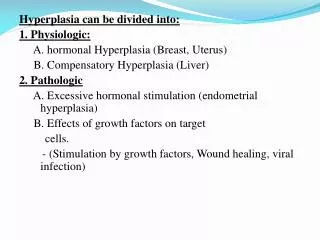
Hyperplasia can be divided into: 1. Physiologic: A. hormonal Hyperplasia (Breast, Uterus) B. Compensatory Hyperplasia (Liver) 2. Pathologic A. Excessive hormonal stimulation (endometrial hyperplasia) B. Effects of growth factors on target cells.

E N D
Hyperplasia can be divided into: 1. Physiologic: A. hormonal Hyperplasia (Breast, Uterus) B. Compensatory Hyperplasia (Liver) 2. Pathologic A. Excessive hormonal stimulation (endometrial hyperplasia) B. Effects of growth factors on target cells. - (Stimulation by growth factors, Wound healing, viral infection)
Cell injury • cell injury results when cells are stressed so severely that they are no longer able to adapt or when cells are exposed to intrinsically damaging agents or suffer from intrinsic abnormalities. • Types of cell injury • 1-reversible • 2-irreversible
Reversible cell injury • In early stages or with mild forms of injury the functional and morphologic changes are reversible if the damaging stimulus is removed. • At this stage the injury has typically not progressed to severe membrane damage and nuclear dissolution. • Types: 1-Cellular swelling 2-Fatty change.
Cellular swelling • Microscopic examination may reveal small, clear vacuoles within the cytoplasm; these represent distended and pinched-off segments of the ER. • This pattern of nonlethal injury is sometimes called hydropic change or vacuolar degeneration
Fatty change -Abnormal accumulation of fat within parenchymal cell. - It occurs in cells participating in fat metabolism e.ghepatocytes and myocytes - manifested by the appearance of lipid vacuoles in the cytoplasm Cause of fatty change: Alcohol; Malnutrition; Diabetes mellitus; Obesity Hepatoxins; Chronic illnesses
The ultrastructural changes of reversible cell injury • include: • (1) plasma membrane alterations such as blebbing, blunting or distortion of microvilli, and loosening of intercellular attachments • (2) mitochondrial changes such as swelling and the appearance of phospholipid-rich amorphous densities • (3) dilation of the ER with detachment of ribosomes and dissociation of polysomes • (4) nuclear alterations, with clumping of chromatin
Subcellular Alterations in Cell Injury: Effects of Injurious Agents on Organelles and Cellular Components • Some forms of cell injury affect particular organelles and have unique manifestations. • Autophagy: In nutrient-deprived cells, organelles are enclosed in vacuoles that fuse with lysosomes. In some cases indigestible pigment (e.g. lipofuscin) remains. • Hypertrophy of SER: Cells exposed to toxins that are metabolized in the SER show hypertrophy of the ER, a compensatory mechanism to maximize removal of the toxins. • Mitochondrialalterations: Changes in the number, size, and shape of mitochondria are seen in diverse adaptations and responses to chronic injury. • Cytoskeletalalterations: Some drugs and toxins interfere with the assembly and functions of cytoskeletal filaments or result in abnormal accumulations of filaments.
A normal cell and the changes in reversible and irreversible cell injury (necrosis)
IrreversibleInjury: OVERVIEW OF CELL INJURY AND CELL DEATH Apoptosis & Necrosis
Two phenomena consistently characterize irreversibility: • 1- The inability to reverse mitochondrial dysfunction (lack of oxidative phosphorylation and ATP generation) even after resolution of the original injury • 2- Profound disturbances in membrane function. As mentioned earlier, injury to lysosomal membranes results in the enzymatic dissolution of the injured cell that is characteristic of necrosis.
Apoptosis • When a cell is deprived of growth factors or the cell's DNA or proteins are damaged beyond repair, the cell kills itself by another type of death, called apoptosis, which is characterized by nuclear dissolution without complete loss of membrane integrity. • Apoptosis is an active, energy-dependent, tightly regulated type of cell death that is seen in some specific situations • apoptosis serves many normal functions and is not necessarily associated with pathologic cell injury
Necrosis = Cell death • Necrosis = morphologic changes that follow cell death in a living tissue or organ resulting from the progressive degradation of enzymes on lethally injured cells. • Necrotic cells are unable to maintain membrane integrity, and their contents often leak out. • The enzymes responsible for digestion of the cell are derived either from the lysosomes of the dying cells themselves or from the lysosomes of leukocytes that are recruited as part of the inflammatory reaction to the dead cells.
Morphology of necrosis • 1- Increased eosinophilia (i.e., pink staining from the eosin dye, the "E" in "H&E"). This is attributable in part to increased binding of eosin to denatured cytoplasmic proteins and in part to loss of the basophilia that is normally imparted by the ribonucleic acid (RNA) in the cytoplasm • 2- Dead cells may be replaced by large, whorled phospholipid masses, called myelin figures, that are derived from damaged cellular membranes • 3- Calcification of such fatty acid residues results in the generation of calcium soaps. Thus, the dead cells may ultimately become calcified.
4- discontinuities in plasma and organelle membranes • 5- marked dilation of mitochondria with the appearance of large amorphousdensities • 6- disruptionoflysosomes • 7- profound nuclear changes culminating in nucleardissolution:
Nuclear dissolution assumes one of 3 patterns • Karyolysis: The basophilia of the chromatin may fade (due to deoxyribonuclease (DNase) activity. • Pyknosis. characterized by nuclear shrinkage and increased basophilia; the DNA condenses into a solid shrunken mass. • Karyorrhexis. the pyknotic nucleus undergoes fragmentation. • In 1 to 2 days, the nucleus in a dead cell completely disappears
Patterns of Tissue Necrosis 1: Coagulative necrosis • Most common • There is loss of nuclei with preservation of the basic cellular shape permitting the recognition of the cell outline and tissue architecture. • Caused by sudden ischemia resulting in denaturation of proteins and enzymes which block proteolysis of the cells. • Infarcts of solid organs except the brain.
2: Liquefactivenecrosis • liquefaction completely digests the dead cells resulting in transformation of the tissue into a liquid viscous mass • If the process was initiated by acute inflammation the necrotic material is frequently creamy yellow and is called pus • Examples: 1. Ischemic destruction of brain. 2. Bacterial lesions ( Abscesses)
3: Caseousnecrosis • The term "caseous" (cheese-like) is derived from the friable yellow-white appearance of the area of necrosis • Coagulative necrosis + liquefaction • Cells do not retain their outlines and do not disappear by lysis. • Very significant example:Tuberculosis • Caseous necrosis is often enclosed within a distinctive inflammatory border composed of histiocytes & lymphocytes known as a granuloma
4: Fatnecrosis • Occurs in adipose tissue • Due to the action of lipases • Commonly seen in acutepancreatitis or due to trauma. • pancreatic enzymes that have leaked out liquefy the membranes of fat cells in the peritoneum • The released fatty acids combine with calcium to produce grossly visible chalky white areas (fat saponification)
5: Gangrenousnecrosis • Ischemic death and coagulative necrosis involving multiple tissue layers. • Generally applied to lower limbs • Types: 1- Dry Gangrene 2- Wet Gangrene modified by liquefactive action of bacteria and leukocytes (superimposed infection)
6- Fibrinoidnecrosis • is a special form of necrosis usually seen in immune reactions involving bloodvessels. • Result from complexes of antigens and antibodies are deposited in the walls of arteries. Deposits of these "immunecomplexes," together with fibrin that has leaked out of vessels, result in a bright pink and amorphous appearance in H&E stains, called "fibrinoid" (fibrin-like) by pathologists. • examples are immunologically mediated vasculitis (like polyarteritis nodosa)
Liquefactive necrosis Caseous necrosis Liquefactive necrosis
Fat necrosis Fibrinoid necrosis Fat necrosis
The small intestine is infarcted. The dark red to grey infarcted bowel contrasts with the pale pink normal bowel at the bottom.
Gangrene, or necrosis of many tissues in a body part, the toes were involved in a frostbite injury. This is an example of "dry" gangrene in which there is mainly coagulative necrosis from the anoxic injury.
Gangrene of the lower extremity. In this case the term "wet" gangrene is more applicable because of the liquefactive component from superimposed infection in addition to the coagulative necrosis from loss of blood supply. This patient had diabetes mellitus.