Download
1 / 68
700 likes | 1.12k Vues
Anatomy, physiology and pathology of the respiratory system (“but mainly the lungs”). Dr Andrew Potter Registrar Department of Radiation Oncology Royal Adelaide Hospital. Anatomy. Overview. Consists of nose, pharynx, larynx, trachea, bronchi, lungs

E N D
Anatomy, physiology and pathology of the respiratory system (“but mainly the lungs”) Dr Andrew Potter Registrar Department of Radiation Oncology Royal Adelaide Hospital
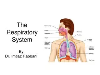
Overview • Consists of nose, pharynx, larynx, trachea, bronchi, lungs • Conducting portion and respiratory portion • Obtains O2 and eliminates CO2 to external environment • Helps regulate pH by adjusting rate of removal of acid-forming CO2
Nose • External portion • Bone and cartilage, covered by skin • Mucous membrane lining • Nostrils, midline septum • Internal portion • Skull cavity inferior to cranium, superior to mouth • Bounded by ethmoids, maxillae, palatine bone, inferior nasal conchae • Communicates with pharynx through the choanae • Communicates with paranasal sinuses • frontal, sphenoid, maxillary, ethmoid • Openings of naso-lacrimal ducts, Eustachian tubes
Pharynx (throat) • Funnel-shaped tube, ~13cm long • Starts at choanae (internal nares) extending to level of cricoid cartilage • Posterior to nasal cavity, oral cavity, larynx • Anterior to cervical vertebral bodies • Muscular wall lined by mucous membrane
Nasopharynx • Uppermost part of pharynx • Posterior to nasal cavity • Extends to plane of soft palate • Eustachian tube openings • Allows air exchange to equalise ear/nose/throat pressures • Pharyngeal tonsils (adenoids) on post wall
Oropharynx • Posterior to oral cavity • Extends from soft palate to level of hyoid • Common passage way for air, food, fluid - communicates with oral cavity • Palatine and lingual tonsils
Hypopharynx (laryngopharynx) • Extends downwards from hyoid • Continuous with oesophagus (posteriorly) and larynx (anteriorly) • Common passage way for air and food
Larynx (voice box) • Connects pharynx with trachea • Epiglottis • cartilage valve to separate food and air • Midline in neck, anterior to C4-C6 • Wall consists of 9 pieces of cartilage • 3 single • Thyroid, epiglottis and cricoid • 3 paired • Arytenoid, corniculate, cuneiform • Vocal cords - false (ventricular) and true • Vibration of vocal cords results in phonation • Barrier against foreign bodies entering lower respiratory tract
Upper respiratory tract - summary • System of interconnected spaces • Transports, filters, humidifies and warms inspired air • Receptors for smell in the nasal cavity • Paranasal sinuses act as resonating chambers for speech • Also reduce weight of facial skeleton
Trachea • Tubular air passage way ~12cm long, 2.5cm diameter • Anterior to oesophagus • Extends from larynx (cricoid cartilage) to ~T5 • Bifurcation at T5 (carina)into left and right main bronchi
Trachea • 16-20 incomplete ‘C’-shaped hyaline cartilage rings provide rigidity • Open part of each ring faces posteriorly to oesophagus • Allows for oesophageal expansion during swallowing • Transverse smooth muscle (trachealis) and elastic connective tissue attach open ends of cartilage rings
Trachea • Important relations • Anteriorly: thyroid isthmus, inferior thyroid veins, sternohyoid and sternothyroid muscles, manubrium, thymus remnants • Laterally: lobe of thyroid, carotid sheath, SVC (right), aortic arch and branches (left), • Posteriorly: oesophagus, recurrent laryngeal nerves
Trachea • Ciliated pseudo-stratified columnar epithelium • Seromucous glands and ducts • humidify air • Cilia (‘brush border’) • Transport excess mucus, foreign bodies upwards like an escalator
Primary (main) bronchi • Incomplete cartilage rings • Stratified columnar epithelium as in trachea • Right main bronchus • To right lung • Shorter, wider and more vertical than left • More prone to foreign bodies lodging • Left main bronchus • To left lung
Secondary (lobar) bronchi • One for each lobe of each lung • 2 on the left • 3 on the right • Further division into tertiary (segmental) bronchi to supply each segment of each lobe • …progressive branching until reaching bronchioles and finally terminal bronchioles and alveolar ducts
Structural features • Gradual transition from one type of airway to the next • Epithelium • Tall, pseudostratified columnar ciliated epithelium in larynx and trachea • Simple cuboidal non-ciliated in small airways • Goblet cells (mucus secreting) gradually disappear
Structural features • Lymphoid aggregates (MALT) • Produces IgA antibodies secreted onto mucosal surface • protection against invading micro-organisms • Smooth muscle • Lies deep to mucosa (except in trachea) • Becomes increasingly important as airway diameter decreases • Regulates calibre of airway and hence resistance to air flow • Sympathetic - muscle relaxation • Parasympathetic - constriction
Structural features • Serous and mucous glands • Progressively less numerous in narrower airways • Cartilage • Supporting skeleton for larynx, trachea and bronchi • Maintains patency during respiration • Gradually diminishes; absent beyond tertiary bronchi
Lungs - gross anatomy • Paired, cone-shaped organs in thoracic cavity • Separated by heart and other mediastinal structures • Covered by pleura • Fibrous membrane with overlying flattened epithelium • Outer layer - parietal pleura, attached to chest wall • Inner layer - visceral pleural, attached to lung surface • Potential space between the two layers (pleural cavity) • Normally contains small amount of pleural fluid - reduces friction between surfaces during movement of respiration
Lungs - gross anatomy • Extend from diaphragm inferiorly to just above clavicles superiorly • Lies against thoracic cage (pleura, muscles, ribs) anteriorly, laterally and posteriorly • Inferior lung base is concave and fits over convexity of each hemi-diaphragm • Narrow superior apex • Surface curved to match curvature of rib cage
Lungs - gross anatomy • Hilum • Medial ‘root’ of the lung • Point at which vessels, airways and lymphatics enter and exit • Cardiac notch • Lies in medial part of left lung to accommodate the heart
Lobes and fissures • Lungs divided into lobes by fissures • Both have an oblique fissure extending forwards and downwards • Separates upper and lower lobes on left • Separates upper, middle and lower lobes on right • Right lung also has horizontal fissure • Separates upper and middle lobes • Each lobe has its own secondary (lobar) bronchus • Named according to the lobe supplied • Further subdivision of each lobe into segments • …similarly supplied by a tertiary (segmental) bronchus
Lobules • Each segment has multiple small compartments - lobules • Each wrapped in connective tissue • Contains lymphatic vessel, arteriole, venule, branch from terminal bronchiole • Terminal bronchioles subdivide into microscopic respiratory bronchioles
Alveoli • Cup-shaped outpouchings • Clustered in alveolar sacs • Resemble microscopic bunches of grapes • Lined by epithelium • Thin elastic basement membrane • Lined by type I alveolar cells with occasional type II alveolar cells • Type II cells secrete alveolar fluid and surfactant • Surfactant acts to reduce surface tension of alveolar fluid (like detergent), helping to keep alveoli from snapping shut
Alveoli • Alveolar macrophages (dust cells) • Phagocytes that remove dust and debris from alveolar spaces • Derived from peripheral blood monocytes • Alveoli surrounded by capillary network to facilitate gas exchange • Single layer of endothelium and basement membrane
Alveolar-capillary membrane • Diffusion of gas between air and circulation occurs across alveolar and capillary walls • Type I and II alveolar cells • Epithelial basement membrane beneath alveolar wall • Capillary basement membrane • Capillary endothelium • Total thickness ~0.5µm • Approx 300 million alveoli in normal lung • Results in large surface area (~70m2) for gas exchange
Lung - blood supply • Dual supply • Bronchial supply • Bronchial arteries supply bronchi, airway airway walls and pleura • Pulmonary supply • Pulmonary arteries enter at hila and branch with airways • Deoxygenated blood from right ventricle pulmonary trunk left and right pulmonary arteries arterioles capillaries oxygenated blood tovenules pulmonary veins left atrium • Venous return is common (ie. both return via pulmonary veins)
Lymphatics • Lymphatic drainage follows vessels • Parabronchial (peribronchial) lymphatics and nodes hilar nodes mediastinal nodes pre- and para-tracheal nodes supraclavicular nodes
Mechanics of breathing • Inspiration - an active process • Diaphragm lowers • Ribs pivot upwards • Intercostal muscles contract • Action similar to a swinging bucket handle • Intra-thoracic pressure lowers • Intrapleural pressure is normally 4mmHg lower than atmospheric pressure, ‘sucking’ the lungs outwards • Lung expands • As volume increases, pressure decreases - Boyle’s law • Air flows from higher atmospheric pressure (760mmHg) into low pressure of the lungs (758mmHg)
Mechanics of breathing • Expiration - passive • Inspiratory muscles relax • Ribs move downwards • Diaphragm relaxes and its domes rise • Surface tension of alveolar fluid causes an inward pull • Elastic recoil of alveolar basement membranes • Reverse pressure gradient • 762mmHg in lungs, 760mmHg atmospheric • Gas pushed out
Respiration • External (pulmonary) respiration • exchange of O2 and CO2 between respiratory surfaces and the blood (breathing) • Internal respiration • exchange of O2 and CO2 between the blood and cells • Cellular respiration • process by which cells use O2 to produce ATP
External respiration • Exchange of O2 and CO2 between alveoli and blood • Partial pressure of O2 higher in alveoli (105mmHg) than blood (40mmHg) so O2 diffuses into blood • Partial pressure of CO2 higher in blood (45mmHg) than alveoli (40mmHg), so CO2 moves into alveoli in opposite direction and gets exhaled out
Internal respiration • Exchange of O2 and CO2 between blood and tissues • Pressure of O2 higher in blood than tissues so O2 gets release into tissues. • Pressure of CO2 higher in tissue than in blood so CO2 diffused in opposite direction into blood. • CO2 is a waste product • O2 is used in cellular respiration
Pulmonary respiration • Internal respiration
Gas transport in blood • Carbon dioxide • 70% as bicarbonate ion (HCO3-) dissolved in plasma • 23% bound to hemoglobin • 7% as CO2 dissolved in plasma • Oxygen • 99% bound to hemoglobin • 1% as O2 dissolved in plasma
Control of breathing • Respiratory centre in reticular formation of the brain stem • Medullary rhythmicity centre • Controls basic rhythm of respiration • Inspiratory (predominantly active) and expiratory (usually inactive in quiet respiration) neurones • Drives muscles of respiration • Pneumotaxic area • Inhibits inspiratory area • Apneustic area • Stimulates inspiratory area, prolonging inspiration
Regulation of respiratory centre • Chemical regulation • Most important • Central and peripheral chemoreceptors • Most important factor is CO2 (and pH) • in arterial CO2 causes in acidity of cerebrospinal fluid (CSF) • in CSF acidity is detected by pH sensors in medulla • medulla rate and depth of breathing