Download
1 / 106
1.17k likes | 2.07k Vues
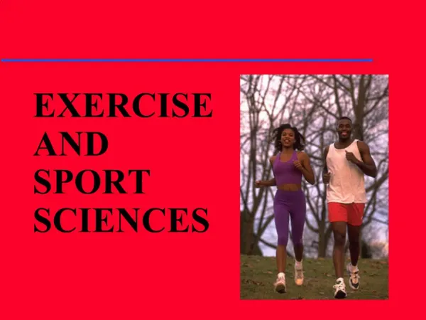
Functional Human Physiology for the Exercise and Sport Sciences The Respiratory System. Jennifer L. Doherty, MS, ATC Department of Health, Physical Education, and Recreation Florida International University. Overview of Respiratory Function. Respiration = the process of gas exchange

E N D
Functional Human Physiologyfor the Exercise and Sport Sciences The Respiratory System Jennifer L. Doherty, MS, ATC Department of Health, Physical Education, and Recreation Florida International University
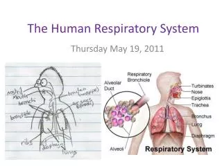
Overview of Respiratory Function Respiration = the process of gas exchange • Two levels of respiration: • Internal respiration (cellular respiration) • The use of O2 with mitochondria to generate ATP by oxidative phosphorylation • CO2 is the waste product • External respiration (ventilation) • The exchange of O2 and CO2 between the atmosphere and body tissues
Internal respiration (cellular respiration) • Involves gas exchange between capillaries and body tissues cells • Tissue cells continuously use O2 and produce CO2 during metabolism • Partial pressure (P) • The PO2 is always higher in arterial blood than in the tissues • The PCO2 is always higher in the tissues than in arterial blood • O2 and CO2 move down their partial pressure gradients • O2 moves out of the capillary into the tissues • CO2 moves out of the tissues into the capillary
External respiration (ventilation) 4 Processes: • Pulmonary Ventilation • Movement of air into the lungs (inspiration) and out of the lungs (expiration) • Exchange of O2 and CO2 between lung air spaces and blood • Transportation of O2 and CO2 between the lungs and body tissues • Exchange of O2 and CO2 between the blood and tissues
Overview of Pulmonary Circulation Deoxygenated blood • Under resting conditions, 5 liters of deoxygenated blood are pumped to the lungs each minute from the right ventricle • CO2 blood concentration is higher than O2 blood concentration in: • Systemic veins • Right atrium • Right ventricle • Pulmonary arteries
Overview of Pulmonary Circulation Oxygenated blood • Transported from the pulmonary capillaries → pulmonary veins → left atrium → left ventricle → aorta → systemic arterial circulation • O2 blood concentration is higher than CO2 blood concentration in: • Alveoli • Pulmonary capillaries • Pulmonary veins • Left atrium • Left ventricle • Systemic arteries
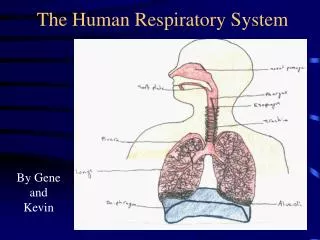
Anatomy of the Respiratory Zone • Gas exchange occurs between the air and the blood within the alveoli
Anatomy of the Respiratory Zone • Alveoli (singular is alveolus) • Tiny air sacs clustered at the distal ends of the alveolar ducts • Alveoli have a thin respiratory membrane separating the air from blood in pulmonary capillaries
Respiratory Membrane The thin alveolar wall consists of: • The fused alveolar and capillary walls • Alveolar epithelial cells • Capillary endothelial cells • The basement membrane • Sandwiched between the alveolar epithelial cells and the endothelial cells of the capillary
Respiratory Membrane • Gas exchanges occurs across the respiratory membrane • It is < 0.1 μm thick • Lends to very efficient diffusion • It is the site of external respiration and diffusion of gases between the inhaled air and the blood • Occurs in the pulmonary capillaries
Structures of the Thoracic Cavity • A container with a single opening, the trachea • Volume of the container changes • Diaphragm moves up and down • Muscles move the rib cage in and out • Volume of the thoracic cavity increases by enlarging all diameters • ↑ diameter = ↑ volume
Boyle’s Law • Volume and pressure are inversely related • ↑ volume = ↓ pressure • Air always flows from an area of higher pressure to an area of lower pressure • Decreased pressure in the thoracic cavity in relation to atmospheric pressure causes air to flow into the lungs • The process of inspiration
Structures of the Thoracic Cavity • Pleura • Parietal pleura: A membrane that lines the interior surface of the chest wall • Visceral pleura: A membrane that lines the exterior surface of the lungs • Intrapleural space • A thin compartment between the two pleurae filled with intrapleural fluid
Pulmonary Pressures • Pressure gradient • The difference between intrapulmonary and atmospheric pressures • 4 Pulmonary Pressures • Atmospheric pressure • Intra-alveolar (Intrapulmonary) pressure • Intrapleural pressure • Transpulmonary pressure
Pulmonary Pressures Atmospheric pressure • The pressure exerted by the weight of the air in the atmosphere (~ 760 mmHg at sea level) Intra-alveolar (Intrapulmonary) pressure • The pressure inside the lungs Intrapleural pressure • The pressure inside the pleural space Transpulmonary pressure • The difference between the intrapleural and intra-alveolar pressure
Pleural Pressures • Intrapleural pressure • The pressure inside the pleural space or cavity • This cavity contains intrapleural fluid, necessary for surface tension • Surface tension • The force that holds moist membranes together due to an attraction that water molecules have for one another • Responsible for keeping lungs patent
Surface Tension • The force of attraction between liquid molecules • Type II alveolar cells secrete surfactant • Creates a thin fluid film in the alveoli • Surfactant (a phospholipoprotein) reduces the surface tension in the alveoli • It interferes with the attraction between fluid molecules • Decreasing surface tension reduces the amount of energy required to expand the lungs
Inspiration • Drawing or pulling air into the lungs • Atmospheric pressure forces air into the lungs • The diaphragm moves downward, decreasing intra-alveolar pressure • The thoracic rib cage moves upward and outward, increasing the volume of the thoracic cavity • Surface tension • Holds the pleural membranes together, which assists with lung expansion • Surfactant reduces surface tension within the alveoli
Inspiration • During inspiration, forces are generated that attempt to pull the lungs away from the thoracic wall • Surface tension of the intraplueral fluid hold the lungs against the thoracic wall, preventing collapse
Expiration • Pushing air out of the lungs • Results due to the elastic recoil of tissues and due to the surface tension within the alveoli • Expiration can be aided by: • Thoracic and abdominal wall muscles that pull the thoracic cage downward and inward, decreasing intra-alveolar pressure • This compresses the abdominal organs upward and inward, decreasing the volume of the thoracic cavity
Muscles of Breathing - Inspiration Quiet Breathing • Muscles include: • External intercostals • Diaphragm • Contract to expand the rib cage and stretch the lungs = ↑ volume of the thoracic cavity • ↑ intrapulmonary volume • ↓ intrapulmonary pressure (relative to atmospheric pressure) • Decreased pressure inside the lungs pulls air into the lungs down its pressure gradient until intrapulmonary pressure equals atmospheric pressure
Muscles of Breathing - Inspiration Forced or Deep Inspiration • Involves several accessory muscles: • Sternocleidomastoid • Pectoralis minor • Scalenes (neck muscles) • Maximal ↑ in thoracic volume • Greater ↓ in intrapulmonary pressure • More air moves into the lungs • At the end of inspiration, the intrapulmonary pressure equals atmospheric pressure
Muscles of Breathing - Expiration Quiet Breathing • Passive process • Depends on the elasticity of the lungs • Muscles of inspiration relax • The rib cage descends • The lungs recoil • ↓ intrapulmonary volume • ↑ intrapulmonary pressure • Alveoli are compressed, thus forcing air out of the lungs
Muscles of Breathing - Expiration Forced Expiration • It is an active process • Occurs in activities such as blowing up a balloon, exercising, or yelling • Abdominal wall muscles are involved in forced expiration • Function to ↑ the pressure in the abdominal cavity forcing the abdominal organs upward against the diaphragm • ↓ volume of the thoracic cavity • ↑ pressure in the thoracic cavity • Air is forced out of the lungs
Factors Affecting Pulmonary Ventilation Lung compliance • The ease with which the lungs may be expanded, stretched, or inflated • Depends primarily on the elasticity of the lung tissue • Elasticity refers to the ability of the lung to recoil after it has been inflated
Factors Affecting Pulmonary Ventilation • Lung and thoracic cavity tissue has a natural tendency to recoil, or become smaller • Lung recoil is essential for normal expiration and depends on the fibroelastic qualities of lung tissue • In normal lungs there is a balance between compliance and elasticity
Factors Affecting Pulmonary Ventilation • Recoil pressure is inversely proportional to compliance • Increased compliance results in decreased recoil • Example: Emphysema • Results in difficulty resuming the shape of the lung during exhalation • Decreased compliance results in increased recoil • Example: Cysitc fibrosis • Results in difficulty expanding the lung because of increased fibrous tissue and mucous
Factors Affecting Pulmonary Ventilation Airway Resistance • Opposition to air flow in the respiratory passageways • Resistance and air flow are inversely related • ↑ airway resistance = ↓ air flow (and vice versa) • Airway resistance is most affected by changes in the diameter of the bronchioles • ↓ diameter of the bronchioles = ↑ airway resistance • Examples: • Asthma • Bronchiospasm during an allergic reaction • A high resistance to air flow produces a greater energy cost of breathing
The Respiratory System: Gas Exchange and Regulation of Breathing Jennifer L. Doherty, MS, ATC Department of Health, Physical Education, and Recreation Florida International University
Diffusion of Gases Partial Pressure of Gases (Pgas) • Concentration of gases in a mixture (air) • Gases move from areas of high partial pressure to areas of low partial pressure • Movement of gases also occurs between cells and the blood in the capillaries • Movement of gases occurs between blood in the pulmonary capillaries and the air within the alveoli • Movement of gasses is by diffusion across the respiratory membrane of the alveoli