Download

1 / 116
1.19k likes | 1.55k Vues
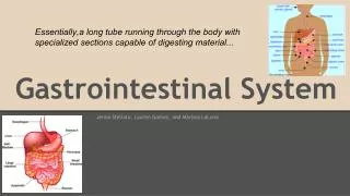
Introduction to the Gastrointestinal System. Summary. Anatomy & Physiology, Pathology and Operative Considerations for: GI System Breast IVAD Care & Use of Endoscopes. Gastrointestinal Definitions.

E N D
Summary • Anatomy & Physiology, Pathology and Operative Considerations for: • GI System • Breast • IVAD • Care & Use of Endoscopes
Gastrointestinal Definitions • Adhesion- tissue that is normally separate is bound together; produced by inflammation, injury, or intentionally surgically created • Anastomosis- joining of parts to create a union • Bile- yellow-green alkaline fluid produced by the liver that aids in digestion and fat absorption • Biliary tract- system of the body involved with bile production, secretion, and transport • Cholangiogram- injection of contrast media into the cystic duct or a tube placed in the common bile duct to allow visualization of the biliary ductal system • Cholecystitis- inflammation of the gallbladder • Cholelithiasis- stones in the gallbladder • Colon- large intestines
Gastrointestinal Definitions • Diverticula- small pouches in the lining or wall of a canal or organ, most commonly the colon • Dysphagia- difficulty swallowing • Fissure- crack or opening • Fistula- abnormal passage between two surfaces or two hollow organs • Intussuseption- when part of the upper intestine slips into or invaginates into a lower portion of the intestine/creates an intestinal obstruction • Meckel’s diverticulum- congenital blind pouch usually associated with the ileum and ileocecal valve • Mucosa- mucous membrane
Gastrointestinal Definitions • Peptic ulcer- open lesion in the stomach or duodenum • Peritonitis- inflammation of the peritoneal cavity • Polyp- growth or tumor with a stalk or pedicle extending from a mucous membrane • Pyloric stenosis- congenital narrowing between the stomach and duodenum (pyloric orifice) due to thickening of circular muscle surrounding it • Resection- excision of a structure and reconstruction of what remains • Sphincter- ring-like muscle surrounding an orifice • Volvulus- twisting or torsion of the intestine causes obstruction and possible strangulation
General Surgery • Abdominal Wall • Abdominal Cavity • Abdominal Organs • Breast (excluding reconstructive procedures) • Vascular Access (excluding dialysis shunting access procedures) • Can include tracheotomy, thyroidectomy and parathyroidectomy
Anatomy of the Abdominal Wall • Subcuticular (skin) • Subcutaneous (fatty/adipose layer) • Anterior fascia (thin or thick membrane over the muscle) • Muscle • Posterior fascia (thin or thick membrane under the muscle) • Peritoneum (shiny membrane covering the abdominal cavity) • Omentum (lesser and greater) • Contents of abdominal cavity (organs/viscera)
Abdominal Cavity • Diaphragm to pelvic base • Pelvic girdle • Ribs • Vertebrae
Abdominal Surgery Landmarks • Xiphoid process • Subcostals • Iliac crests • Symphysis pubis • Umbilicus • Linea alba • Serve as reference points for incisions and internal organ access
Abdominal Divisions • Four Quadrants • Nine Quadrants
Abdominal Division • Anatomy of the Abdomen RUQ (right upper quadrant) contents: liver gallbladder duodenum head of pancreas right kidney and adrenal part of ascending and transverse colon
Anatomy of Abdomen Continued • LUQ (left upper quadrant) contents: stomach spleen left lobe of liver body of pancreas left kidney and adrenal part of transverse and descending colon
Anatomy of Abdomen Continued • RLQ (right lower quadrant) contents: cecum appendix right ovary and fallopian tube right ureter right spermatic cord
Anatomy of Abdomen Continued • LLQ (left lower quadrant) contents: part of descending colon sigmoid colon left ovary and fallopian tube left ureter left spermatic cord
Anatomy of Abdomen Continued • Midline of Abdomen: Aorta Uterus Bladder
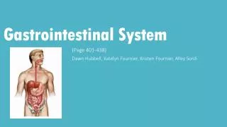
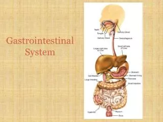
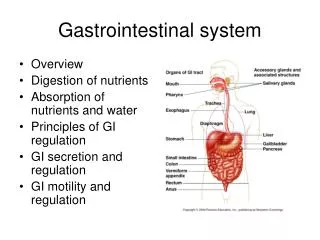
Digestive TractAlimentary Canal • Mouth to the Anus • Mouth>Pharynx>Pharyngoesophageal Sphincter>Esophagus> Esophagogastric Sphincter>Stomach>Pyloric Sphincter >Duodenum>Jejunum>Ileum>Cecum (appendix)>Ascending Colon>Transverse Colon>Descending Colon>Sigmoid Colon>Rectum>Internal Sphincter>External Sphincter>Anus
Physiology of the Digestive System • Two major parts: • GI tract/alimentary Canal-mouth to anus-about 30 ft long • Accessory Organs-outside of or to side of GI tract, but are connected-teeth, salivary glands, biliary system: liver, gallbladder, pancreas
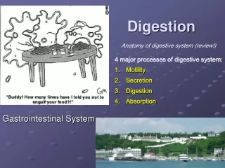
Physiology of the Digestive System • 5 major processes: • Ingestion/eating • Mechanical and Chemical Digestion • Peristalsis • Absorption • Defecation
Mechanical and Chemical Digestion • Begins in mouth, teeth increases surface area of food to allow enzymes to work on • Tongue pushes food underneath teeth and flips food as a “bolus” to back of throat (oropharynx) • Salivary Glands-primary salivary amylase begins break down of carbohydrates
Mechanical and Chemical Digestion • Esophagus: • Begins at oropharynx • Mucous allows food to slide down
Mechanical and Chemical Digestion • Stomach • 4 areas: • Cardiac (esophagus ends and cardiac or esophageal sphincter empties into this region • Fundus/fundic area part that is rounded on left side of body • Body-main part of stomach • Pyloric region or antrum=area before pyloric sphincter which is where the duodenum begins
Mechanical and Chemical Digestion • Rugae (hills and valleys allow stomach to expand • 3 basic cell types here that produce: • Pepsinogen • HCl • Mucous • HCl acid activates pepsinogen which then becomes pepsin which begins protein breakdown • Vagus nerve stimulates tunica muscularis to create waves in stomach from bottom up to allow for mixing of HCl and pepsin • Vagus nerve tires easily, production of hormone gastrin by the stomach sustains the action of stomach wave action • Food in stomach 1-6 hours • Food broken down into “chyme” (semi-solid or pasty material
Pancreas • Head • Body • Tail • 80% comprised of lobules • Lobules consist of exocrine and endocrine glands
Mechanical and Chemical Digestion • Pancreas • Endocrine and exocrine gland 1. Endocrine portion = Islets of Langerhan • No ducts, secrete into blood or lymph • Secreting portion is Islets of Langerhans • 1% of pancreatic mass • Receives 25% pancreatic blood supply
Islets of Langerhan • Two cell types: • Alpha cells secrete hormone glucagon (↑ blood sugar level) • Beta cells secrete hormone insulin (↓ blood sugar levels) • Function maintenance of blood glucose levels
Exocrine glands • Secrete directly through a duct • Called acini • Functions: breakdown fats, proteins, carbohydates and maintain pH • pH maintenance prevents excessive acid production which prevents duodenal ulcers
Mechanical and Chemical Digestion • Pancreas 2. Exocrine portion: • Produces enzymes: collectively called pancreatic juices (Trypsin, chymotrysin, carboxypeptidase) break down proteins • Pancreatic amylase breaks down carbohydrates • Pancreatic lipase breaks down lipids • All get to small intestine via pancreatic duct (Duct of Wirsung) at Ampula of Vater
Mechanical and Chemical Digestion • Liver • Functions: • Store excessive nutrients • Detoxify and filter toxins • Regulate nutrient levels • Destroy worn out RBCs, WBCs, bacteria • Produce heparin, prothrombin, fibrinogen, and albumin • Store fat soluable vitamins (A,D,E,K) • Water soluable are excreted • Produces bile (function to emulsify lipids)
Mechanical and Chemical Digestion • Gallbladder • Stores bile • Sphincter of Oddi (hepatopancreaticsphincter) opens to release bile and pancreatic juices into the small intestine • Bile released from gallbladder when lipids (fats) are present
Mechanical and Chemical Digestion • Small Intestine • Begins at pyloric sphincter, ends at ileocecal valve • About 21 feet long • Where 90% of digestion and absorption occur • Other 10% in stomach and large intestines • 3 parts: • Duodenum-(12 inches long) • Jejunum (8 feet long) • Ileum (12 feet long)
Mechanical and Chemical Digestion • Large intestine • Parts of: Ascending, transverse, descending, sigmoid, rectum • Functions: • Absorption of water, electrolytes, proteins into amino acids, and bacterial products • Feces formation • Food in large intestine 3-10 hours for absorption purposes • Undigested food is expelled via “mass peristaltic movement” out the anus
Esophagus • Esophagitis • Ulceration • Neoplasms • Foreign bodies • Zenker’s diverticulum located in esophagus – dx w/ esophagoscopy - 1°sx dysphagia • Esophageal varices - esophagus erodes due to severe alcoholism
Pathology of The Stomach • Ulcers • Gastritis • Polyps • Bezoar (hairball in animals/fiber ball in humans) • Carcinoma • Lymphoma (benign or malignant)
Small Intestine • Duodenum • Jejunum • Ileum
Pathology of the Small Intestine • Ulcer (duodenum most common site) • Neoplasm (benign or malignant) • Obstruction • Crohn’s Disease (Surgical intervention needed with perforation, abscess or hemorrhagic fistula formation)
Colon Pathology • Appendicitis • Adhesions • Herniation • Polyps • Diverticulosis or Diverticulitis • Tumor (benign or malignant) • Ulcerative Colitis • Obstruction • Volvulus • Intussusception • Impaction
Anorectal Pathology • Fistula • Fissure • Pilonidal Cyst • Hemorrhoids
Pathology of the Pancreas • Cyst • Tumor (Benign or Malignant) • Chronic Pancreatitis • Trauma
Spleen • Largest lymphatic mass in body • Composed of: 75% red pulp (vascular) 25% white pulp (lymphatic/immune response) • Functions: RBC and Plt storage • Excision of renders liver and other lymphatic tissues to pick up the slack
Pathology of the Spleen • Trauma • Hematologic Disorders • Tumor (Benign or Malignant) • Cyst • Splenomegaly