Download

1 / 56
600 likes | 931 Vues
Hemolytic anemia. Rakesh Biswas MD, Professor, Department of Medicine, People's College of Medical Sciences, Bhanpur, Bhopal, India. Young man of 19 Complains of giddiness weakness, pallor Examination reveals a spleen mild lemon yellow sclera.

E N D
Hemolytic anemia Rakesh Biswas MD, Professor, Department of Medicine, People's College of Medical Sciences, Bhanpur, Bhopal, India
Young man of 19Complains of giddiness weakness, pallorExamination reveals a spleenmild lemon yellow sclera
How shall you investigate to find out the cause of the problem?
Laboratory investigations:Severe normochromic, normocytic anemia (hemoglobin level of 6.4 g/dLReticulocyte count of 12.2%. Blood film:
Bilirubin level of 2.5 mg/dL, Lactate dehydrogenase (LDH) of 2140 IU/L,Haptoglobin below 7 mg/dL
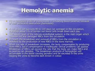
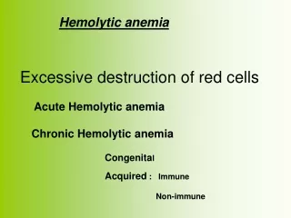
Introduction • Mean life span of a RBC-120days • Removed Extravascularly by- Macrophages of RE system
Hemolytic Anemia • Definition: • Those anemias which result from an increase in RBC destruction • Classification: • Congenital / Hereditary • Acquired
Features of HEMOLYSIS Bilirubin LDH Reticulocytes, n-RBC Haptoglobulins +ve Urinary hemosiderin, Urobilinogen Blood Film Spherocytes No spherocytes Fragmentation DCT +ve DCT –ve AI Hemolysis H. Sherocytosis Malaria, Clostidium Hereditery enzymopathies Microangiopathic, Traumatic
Red Cell Membrane Defects 1.Hereditary Spherocytosis • Usually inherited as AD disorder • Defect: Deficiency of Beta Spectrin or Ankyrin Loss of membrane in Spleen & RES becomes more spherical Destruction in Spleen
C/F: Asymptomatic Fluctuating hemolysis Splenomegaly Pigmented gall stones- 50%
Complications • Clinical course may be complicated with Crisis: • Hemolytic Crisis: associated with infection • Aplastic crisis: associated with Parvovirus infection
Inv: Test will confirm Hemolysis P Smear: Spherocytes Osmotic Fragility: Increased Screen Family members
Management: Folic Acid 5mg weekly, prophylaxis life long Spleenectomy Blood transfusion in Ac, severe hemolytic crisis
2.Hereditary Elliptocytosis • Equatorial Africa, SE Asia • AD / AR • Functional abnormality in one or more anchor proteins in RBC membrane- Alpha spectrin , Protein 4.1 • Usually asymptomatic • Mx: Similar to H. spherocytosis • Variant: 3.SE-Asian ovalocytosis: • Common in Malaysia , Indonesia… • Asymptomatic-usually • Cells oval , rigid ,resist invasion by malarial parasites
Red Cell Enzymopathies • Physiology: • EM pathway: ATP production • HMP shunt pathway: NADPH & Glutathione production
1. Glucose-6-Phosphate Dehydrogenase( G6PD ) Deficiency Pivotal enzyme in HMP Shunt & produces NADPH to protect RBC against oxidative stress Most common enzymopathy -10% world’s population Protection against Malaria X-linked
(Oxidisedform) (Reduced form)
Clinical Features: Acute drug induced hemolysis: Aspirin, primaquine, quinine, chloroquine, dapsone…. Chronic compensated hemolysis Infection/acute illness Neonatal jaundice Favism
Inv: e/o non-spherocytic intravascular hemolyis P. Smear: Bite cells, blister cells, irregular small cells, Heinz bodies, polychromasia G-6-PD level Treatment: Stop the precipitating drug or treat the infection Acute transfusions if required
2. PyruvateKinase Deficiency AR Deficient ATP production, Chronic hemolytic anemia Inv; P. Smear: Prickle cells Decreased enzyme activity Treatment: Transfusion may be required
Autoimmune Hemolytic Anemia • Result from RBC destruction due to RBC autoantibodies: Ig G, M, E, A • Most commonly-idiopathic • Classification • Warm AI hemolysis:Ab binds at 37degree Celsius • Cold AI Hemolysis: Ab binds at 4 degree Celsius
1.Warm AI Hemolysis: Can occurs at all age groups F > M Causes: 50% Idiopathic Rest - secondary causes: 1.Lymphoid neoplasm: CLL, Lymphoma, Myeloma 2.Solid Tumors: Lung, Colon, Kidney, Ovary, Thymoma 3.CTD: SLE,RA 4.Drugs: Alpha methyl DOPA, Penicillin , Quinine, Chloroquine 5.Misc: UC, HIV
MACROCYTE SPHEROCYTE IMMUNOHEMOLYTIC ANEMIA
complement Direct antiglobulin testdemonstrating the presence of autoantibodies (shown here) or complement on the surface of the red blood cell.
Inv: e/o hemolysis, MCV P Smear: Microspherocytosis, n-RBC Confirmation: Coomb’s Test / Antiglobulin test Treatment Correct the underlying cause Prednisolone 1mg/kg po until Hb reaches 10mg/dl then taper slowly and stop Transfusion: for life threatening problems If no response to steroids Spleenectomy or, Immunosuppressive: Azathioprine, Cyclophosphamide
2. Cold AI Hemolysis Usually Ig M Acute or Chronic form Chronic: C/F: Elderly patients Cold , painful & often blue fingers, toes, ears, or nose ( Acrocyanosis) Inv: e/o hemolysis P Smear: Microspherocytosis Ig M with specificity to I or I Ag
Other causes of Cold Agglutination: Infection: Mycoplasma pneumonia, Infec Mononucleosis PCH : Rare cause seen in children in association with cong syphilis
Treatment: Treatment of the underlying cause Keep extremities warm Steroids treatment Blood transfusion
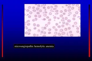
Non-Immune Acquired Hemolytic Anemia 1. Mechanical Trauma A). Mechanical heart valves, Arterial grafts: cause shear stress damage B).March hemoglobinuria: Red cell damage in capillaries of feet C). Thermal injury: burns D). Microangiopathic hemolytic anemia (MAHA): by passage of RBC through fibrin strands deposited in small vessels disruption of RBC eg: DIC,PIH, Malignant HTN,TTP,HUS
Acquired hemolysis 2.Infection F. malaria: intravascular hemolysis: severe called ‘Blackwater fever’ Cl. perfringens septicemia 3.Chemical/Drugs: oxidant denaturation of hemoglobin Eg: Dapsone, sulphasalazine, Arsenic gas, Cu, Nitrates & Nitrobenzene
The direct antiglobulin test was positive for complement (C3d) (++), and IgG (++-). Also was positive for agglutinins of IgM type and had a titer of 1:1024.
Serologies for human immunodeficiency virus, hepatitis B and C viruses, and Mycoplasma pneumoniae were negative. Rheumatoid factor and antinuclear antibodies were undetectable.
Prednisone therapy was started at a dose of 1 mg/kg intravenously, daily. Hemoglobin level rose to 11 g/dL, concomitantly with the improvement of hemolytic signs.
A reduction of positivity of both direct and indirect antiglobulin tests (polyvalent serum + ; C3d + ; IgG+ ), as well as a reduction of cold agglutinin titers (1:128), was observed 8 weeks after corticosteroid therapy.
Three months later, corticosteroids were tapered to a maintenance dose of 25 mg daily. Hemolysis recurred again with the fall of hemoglobin to 7 g/dL.
The direct antiglobulin test recurred positive for polyvalent serum (+++), complement (+++), and IgG (+++), while cold agglutinin titers again became strongly positive (1:256).
Immunophenotyping of bone marrow cells showed that 10% of all the cells were CD20 and CD19 positive.