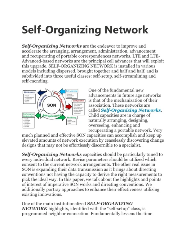
RNA Folding: Structure, Function, and Predictions
Explore the fascinating world of RNA folding and its implications in cellular functions with this lecture on self-assembly in nucleic acids, DNA, and RNA folding. Learn about the primary and secondary structures of RNA, base-pairing, RNA's role in the central dogma, different types of non-coding RNA, RNA structure motifs, and the importance of RNA folding predictions. Discover how RNA folding can predict function and understand the influence of ions on RNA folding kinetics.

RNA Folding: Structure, Function, and Predictions
E N D
Presentation Transcript
Self-Organizing Bio-structures NB2-2009 L.Duroux
Lecture 3 Self-Assembly in nucleicacids
What is RNA? • Aside of being DNA’s “messenger”, RNA performs functions itself • RNA secondary structure is related to mRNA stability & RNA functions • RNA folding can be predicted & the effects of mutations modeled
RNA Primary Structure (-e) Structure of RNA backbone 5' (-e) (-e) (-e) 3' • RNA chain directionality: 5'3' • Backbone carries charge (-e) on each nucleotide • Formation of an RNA structure requires cations
Four Types of Bases Adenine (A) Uracil (U) Guanine (G) Cytosine (C) Purines Pyrimidines
Watson-Crick canonical base pair Base-Pairing: a famous case of molecular self-assembly A U C G
The Central Dogma transcription splicing mRNA tRNA translation ribosome DNA pre mRNA mRNA protein
RNAs are Critical to Cellular Functions • Messenger RNA (mRNA) • codes for protein • Small nuclear RNAs (snRNA) • splice mRNA in nucleus • Transfer RNA (tRNA) • carries amino acid to ribosome • Ribosomal RNA (rRNA) • is the integral part of the ribosome • Small interfering RNA (siRNA) • mRNA turn-over, defense mechanism • Micro RNA (miRNA) • Gene expression regulation
Some biological functions of non-coding RNA • snRNA: RNA splicing, telomere maintenance, transcription regulation • miRNA: translational control (down regulation) • siRNA: RNA interference, gene specific down regulation • Guide RNAs: RNA editing (mitochondria protozoa) • Ribozymes: Catalysis in ribozomes • The function of the RNA molecule depends on its folded structure
The RNA Helix ssRNA forms A-helix: Grooves Binding sites
RNA secondary structure • Defined by base-pairing • Form short helical structures U
Torsion Angles define 3D structure P O c c c O P each bond ~ 1.5 Å nucleotide structure We need 7 torsional angles per nucleotide to specify the 3D structure of an RNA
RNA specific folds • The RNA molecule folds on itself. • The base pairing is as follows: G C A U G U hydrogen bond LOOP U U C G U A A U G C 5’ 3’ 5’ 3’ G A U C U U G A U C STEM
RNA Secondary Structure Motifs Pseudoknot Stem Interior Loop Single-Stranded Bulge Loop Junction (Multiloop) Hairpin loop Image– Wuchty
Secondary structure motifs and symbols Secondary Structure Contact (Base Pair) Tertiary Structure Contact (Base Pair)
RIBOZYMES • Catalytic RNA • Can work alone (Mg2+) or with proteins • Therapeutic applications?
Control of ironlevels by mRNA secondary structure Iron Responsive Element (IRE) on mRNA G U A G CN N N’ N N’ N N’ N N’ C N N’ N N’ N N’ N N’ N N’ conserved Recognized by Iron Responsive Protein (IRP1, IRP2) when Fe deficiency 5’ 3’
Low Iron IRE-IRP inhibits translation of Ferritin IRE-IRP Inhibition of degradation of TR High Iron IRE-IRP off -> Ferritin translated Transferrin receptor degraded F: Ferritin = iron storage TR: Transferrin receptor = iron uptake IRP1/2 IRE 3’ 5’ F mRNA IRP1/2 3’ TR mRNA 5’
Structure-based similarity H H St St I1 I1 B B I2 I2 Sequence Similarity %ID = 34% gurken AAGTAATTTTCGTGCTCTCAACAATTGTCGCCGTCACAGATTGTTGTTCGAGCCGAATCTTACT 64 Ifactor ---TGCACACCTCCCTCGTCACTCTTGATTTT-TCAAGAGCCTTCGATCGAGTAGGTGTGCA-- 58 ** *** ** *** *** * * ***** * * Structural Similarity I Factor : (retrotransposon) 58nt stem loop Gurken : (miRNA controlling development) 64nt stem loop
Goal:To predict function of an RNA from itssequence from: structure stability folding kinetics RNA folding predictions Ultimate goal:To predict RNA function from itssequence
Folding Free Energy of Secondary Structure Folding free energy: ΔG = G ( secondary structure) - G ( ) ΔG = ΔH – T ΔS
Applications for RNA folding predictions • Explain why non-(protein) coding regions are conserved • Viral RNA packing inside capsid • Prediction of functional RNAs • Identify similarity, not by sequence but by structure
Why Study RNA Folding Kinetics? B A conversion is slow as compared with the translational process Conformation B is kinetically trapped. Kinetics is tied to Function
H2O and metal ions are integral parts of nucleic acid structure
[Na+] stabilizes secondary structure From Tinoco & Bustamante,JMB (1999) 273,271 • [Na+] by 10 folds Tm by 3.8 C
Multivalent Ions Stabilize Tertiary Fold Pseudoknot
[Mg2+] Stabilization Na+ = 200mM + 50 From Tinoco & Bustamante,JMB (1999) 273,271
RNA folding kinetics strongly depends on ions Na+ Secondary structure Mg2+ Tertiary structure Metal ion binding sites can be formed before, during, or after the formation of the tertiary structure
Advantages to Double Helix • Stability---protects bases from attack by H2O soluble compounds and H2O itself. • Provides easy mechanism for replication
Formal geometrical models for describing shape of Helix • Allows for molecular modeling based on primary structure • Based on Free-energy computations and minimization algorithms • Useful to predict impact of sequence composition or mutations (non-canonical base-pairing) on helical structure
Parameters that define base pairs 3DNA (v1.5) — A 3-Dimensional Nucleic Acid Structure Analysis and Rebuilding Software Package Xiang-Jun Lu, Wilma K. Olson
Parameters that define sequential base pair steps 3DNA (v1.5) — A 3-Dimensional Nucleic Acid Structure Analysis and Rebuilding Software Package Xiang-Jun Lu, Wilma K. Olson
Parameters that relate base pair to the helical frame 3DNA (v1.5) — A 3-Dimensional Nucleic Acid Structure Analysis and Rebuilding Software Package Xiang-Jun Lu, Wilma K. Olson
Physical Structure (cont’d) • Chains are anti-parallel (i.e in opposite directions) • Diameter and periodicity are consistent • 2.0 nm • 10 bases/ turn • 3.4 nm/ turn • Width consistent because of pyrimidine/purine pairing
G-C Content • A=T, G=C, but AT≠GC • Generally GC~50%, but extremely variable • Examples: • Slime mold~22% • Mycobacterium~73% • Distribution of GC is not uniform in genomes