Download
1 / 71
760 likes | 1.07k Vues
Anatomy of Swallowing. Strucures Muscles Nerves Vascular supply. PALATE. HARD AND SOFT. Forms the arched part of the mouth and floor of nasal cavities. Separates the oral cavity from the nasal cavities and the nasopharynx , part of the pharynx superior to the soft palate.

E N D
Anatomy of Swallowing • Strucures • Muscles • Nerves • Vascular supply
PALATE HARD AND SOFT
Forms the arched part of the mouth and floor of nasal cavities. • Separates the oral cavity from the nasal cavities and the nasopharynx, part of the pharynx superior to the soft palate. • Superior (nasal) surface of the palate is covered with respiratory mucosa and the inferior (oral) surface is covered with oral mucosa densely packed with glands. • The palate consists of two regions: the hard and soft palate posteriorly.
Hard Palate • Palatine processes of the maxillae form the anterior 2/3s of the hard palate. Could be 3/4s • The horizontal plates of the palatine bones form the posterior 1/3. Could be 1/4s • In the oral cavity, the upper alveolar arch borders the hard palate anteriorly and laterally. • Posteriorly the hard palate is continuous with the soft palate.
A single nasal spine is formed at the midline where the 2 horizontal plates join and projects backwards from the margin of the hard palate. The posterior margin of the horizontal plates and the posterior nasal spine are associated with the attachment of the soft palate.
The mucosa of the hard palate in the oral cavity possesses numerous transverse palatine folds ( palatine rugae) and a median longitudinal ridge (palatine raphe) which ends anteriorly in a small oval elevation (incisive papillae) The incisive papillae overlies the incisive fossa formed between the horizontal plates of the maxillae immediately behind the incisor teeth.
GREATER PALATINE FORAMEN • Formed mainly by the horizontal plate of the palatine bone and completed laterally by the adjacent part of the maxilla, opens onto the posterolateral aspect of the horizontal plate. • This foramen is the inferior opening of the palatine canal, which continues superiorly into the pterygopalatinefossa and transmits the greater palatine nerves and vessels to the palate.
Greater palatine foramen medial to the 3rd molar the greater palatine foramen pierces the lateral border of the bony plate • The greater palatine vessels and nerve emerge from this foramen and run anteriorly on the plate • The lesser palatine foramina posterior to the greater palatine foramen pierce the pyramidal process of the palatine bone • These foramina transmit the lesser palatine nerves and vessels to the soft palate and adjacent structures.
Incisive Fossa • Incisive Fossa is a depression in the midline of the bony palate posterior to the central incisive teeth into which the incisive canals open. The nasopalatine nerves pass from the nose though a variable number of incisive canals and foramina that open into the incisive fossa.
incisive fossa Greater palatine foramen Lesser palatine foramen
Innervation Palate • The sensory nerves of the palate are branches of the maxillary nerve ,(CNV2), which branch from the pterygopalatine ganglion. • The greater palatine nerve supplies the gingivae, mucous membrane and glands of most of the hard palate. • The nasopalatine nerve supplies the mucous membrane of the anterior part of the hard palate. • The lesser palatine nerve supply the soft palate.
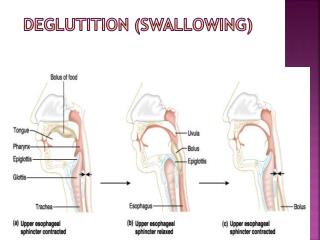
Swallowing • When a person swallows the soft palate initially is tensed to allow the tongue to press against it, squeezing the bolus of food to the back of the mouth. The soft palate is then elevated posteriorly and superiorly against the wall of the pharynx, thereby preventing the passage of food into the nasal cavity.
The soft palate • The structure composed of mucous membranes, muscular fibers, and mucous glands, suspended from the posterior border of the hard palate forming the roof of the mouth. • When the soft palate rises, as in swallowing, it separates the nasal cavity and nasopharynx from the posterior part of the oral cavity and oral portion of the pharynx. • In sucking the soft palate and posterior superior surface of the tongue occlude the oral cavity from the oropharynx, creating a posterior seal. Thus the soft palate prevents the escape of fluid and food up through the nose and with the tongue allows fluid and food to collect in the mouth until swallowed.
UVULA SOFT PALATE HARD PALATE
While swallowing, the soft palate is pushed backwards. This prevents food and drink from entering the nasal cavity; if the soft palate cannot touch the back of the throat while swallowing, food and drink can enter the nasal cavity.
Moveable posterior third of the palate and is suspended from the posterior border of the hard palate. • The soft palate has no bony skeleton however its anterior aponeurotic part is strengthened by the palatine aponeurosis which attaches to the posterior edge of the hard palate. • The aponeurosis is thick anteriorly and thin posteriorly where it blends with a posterior muscular part. • Posterioinferiorly the soft palate has a curved free margin from which hangs the uvula.
Laterally the soft palate is continuous with the wall of the pharynx and joined to the tongue by the palatoglossal and palapharyngeal arches respectively.
Fauces • The Fauces (the throat) is the space between the cavity of the mouth and the pharynx. • The fauces is bounded superiorly by the soft palate and inferiorly by the root of the tongue and laterally by the pillars of the fauces, the palatoglossal and palatopharyngeal arches. • The isthmus of the fauces is the short constricted space that establishes the connection between the oral cavity proper and the oropharynx. The isthmus is bounded anteriorly by the palatoglossal folds and posteriorly by the palatopharyngeal arches. • The palatine tonsils often referred to as the “the tonsils” are masses of lymphoid tissue, one on each side of the oropharynx. • Each tonsil is in a tonsillarfossa (sinus) bounded by the palatoglossal and palatopharyngeal arches and the tongue.
1vestibule 2hard palate 3soft palate 4uvula 5palatoglossal arch 6palatine tonsil 7palatopharyngeal arch 8posterior wall of oropharynx 9pterygoid hamulus
TENSOR VELI PALATINI • Composed of two parts • Vertical muscle muscular part • Horizontal fibrous part which forms the palatine aponeurosis
Vertical part of the tensor velipalatini is thin and triangular in shape with its base attached to the skull and its apex pointed inferiorly. • The base is attached along an oblique line that begins medially at the scaphoid fossa near the root of the pterygoid process of the sphenoid bone and continues laterally along the membranous part of the pharynotympanic tube to the spine of the sphenoid bone.
Tensor VeliPalatini • The tensor velipalatini descends vertically along the lateral surface of the medial plate of the pterygoidprocess and pharyngeal wall to the pterygoidhamulus where the fibers converge to form a small tendon. • The tendon loops 90 degrees medially around the pterygoidhamulus, penetrating the origin of the buccinator muscle as it does and expands like a fan to form the fibrous horizontal part of the muscle. This fibrous part is continuous across the midline with the partner on the other side to form the palatine aponeurosis
Palatine Aponeurosis • Attached anteriorly to the margin of the hard palate but is unattached posteriorly where it ends in a free margin. This expansive aponeurosis is the major structural element of the soft palate to which the other muscles of palate attach.
Tensor VeliPalatini • Actions: • Tenses (makes firm) the soft palate • Opens the mouth of the pharyngotympanic tube (auditory tube) during yawning and swallowing.
The tensor velipalatini it is found lateral to the levator veli palatini muscle. It arises by a flat lamella from the scaphoid fossa at the base of the medial pterygoid plate, from the spina angularis of the sphenoid and from the lateral wall of the cartilage of the auditory tube Descending vertically between the medial pterygoid plate and the medial pterygoid muscle, it ends in a tendon which winds around the pterygoid hamulus, being retained in this situation by some of the fibers of origin of the medial pterygoidmuscle Between the tendon and the hamulus is a small bursa. The tendon then passes medialward and is inserted into the palatine aponeurosis and into the surface behind the transverse ridge on the horizontal part of the palatine bone. • M,, • ,,,,,,,,,,,,,,,,,,,,,,,,,,,,,,,,,,,,,,,,,,,,,,,,,,,,,,,,,,,,,,,,,,,,,,,,,,,,,,,,,,,,,,,,,,,,,,,,,,,nnnnnnnnnnnnnnnnnnnnnnnnnnnnnnnnnnnnnnnnnnnnnnnnnnnnnnnnnnnnnnnnnnnnnnnnnnnnnnnnnnnnnnnnnnnnnnnnnnnnnnnnnnnnnnnnnnnnnnnnnnnnnnnnnnnnnnnnnnnnnnnnnnnnnnnnnnnnnnnnnnnnnnnnnnnnnnnnnnnnnnnnnnnnnnnnnnnnnnnnnnnnnnnnnnnnnnnnnnnnn9999999999999999999999999999999999999o • O9 The tensor velipalatini it is found lateral to the levator veli palatini muscle. It arises by a flat lamella from the scaphoid fossa at the base of the medial pterygoid plate, from the spina angularis of the sphenoid and from the lateral wall of the cartilage of the auditory tube Descending vertically between the medial pterygoid plate and the medial pterygoid muscle, it ends in a tendon which winds around the pterygoid hamulus, being retained in this situation by some of the fibers of origin of the medial pterygoid muscle Between the tendon and the hamulus is a small bursa The tendon then passes medial ward and is inserted into the palatine aponeurosis and into the surface behind the transverse ridge on the horizontal part of the palatine bone.
Levatorvelipalatini • Origin base of skull and descends to the upper surface of the palatine aponeurosis. • On the skull it originates from a roughened area on the petrous part of the temporal bone immediately anterior to the opening of the Carotid canal. • Levatorvelipalatini passes anterioinferiorly through fascia of the pharynotympanic tube and inserts onto the palatine aponeurosis. Its fibers interlaces at the midline with those of the levatorvelipalatini on the other side.
The levatorvelipalatinin does not pass around the pterygoidhamulus but courses directly from the base of the skull to the upper surface of the palatine aponeurosis. • Therefore they elevate the palate above the neutral position and close the pharyngeal isthmus between the nasopharynx and oropharynx.
Palatopharyngeus • Originates from the superior surface of the palatine aponeurosis and passes posterolaterally over its margin to descend and become one of the longitudinal muscles of the pharyngeal wall. • Attached to the palatine aponeurosis by 2 flat lamellae separated by the levatorvelipalatini. • The more anterior and lateral of these 2 lamellae is attached to the posterior margin of the hard palate as well as to the palatine aponeurosis.