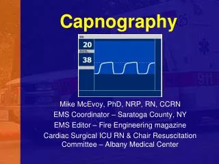
CAPNOGRAPHY
CAPNOGRAPHY . Dr. Radhika Dhanpal radhika.dhanpal@rediffmail.com Professor and Head. Department of Anesthesiology and Critical Care, St. John’s Medical College Hospital Bangalore . Email: anaesthesia.co.in@gmail.com. www.anaesthesia.co.in. ASA


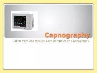
CAPNOGRAPHY
E N D
Presentation Transcript
CAPNOGRAPHY Dr. Radhika Dhanpal radhika.dhanpal@rediffmail.com Professor and Head Department of Anesthesiology and Critical Care,St. John’s Medical College Hospital Bangalore Email: anaesthesia.co.in@gmail.com www.anaesthesia.co.in
ASA House of delegates July 1, 2011 implementation of Oct 2010 decision “Standards for Basic Anesthesia Monitoring”During regional anesthesia (with no sedation ) or local anesthesia (with no sedation ), the adequacy of ventilation shall be evaluated by continual observation of qualitative clinical signs. During moderate or deep sedation, the adequacy of ventilation shall be evaluated by continual observation of qualitative clinical signs and monitoring for the presence of exhaled CO2 unless precluded or invalidated by the nature of the patient, procedure or equipment” ISA – Desirable standard 1999
Definition :Graphic display of instantaneous CO2 concentration • Luft • Collier • Ramwell • Holland in 1978 , was the first country to adopt it as a standard of monitoring during anaesthesia .
Methods of measurement • Infrared spectrography • Raman spectrography • Mass spectrography • Photoacoustic spectrography • Chemical colorimetric analysis
Raman spectrography Gas sample is aspirated into the analysing chamber where it is illuminated by a high intensity monochromatic argon laser beam. The light is absorbed by molecules which are then excited to unstable vibrational or rotational energy states, these Raman scattering signals are then measured.
Mass spectrography It separates gases and vapors of different molecular weight on the basis of their mass into a spectrum. By analyzing the spectrum, the composition and relative abundance of each gas in a sample can be determined .
Infrared method : Infrared waves at 4.3 mm are absorbed by certain gases producing absorption bands on the infrared electromagnetic spectrum.
Photoacoustic gas measurements The gas to be measured is irradiated by modulated light of a pre-selected wavelength . The light beam when chopped, generates an acoustic signal which is detected by two microphones.
Colorimetric method Chemically treated foam indicator attached to endotracheal tube.
Factors influencing the reading ; • Atmospheric pressure : Changes in atmospheric pressure are usually of the order of 20 mm Hg . This results in a change in PaCO2 of less than 0.5 - 0.8 mm Hg • PEEP . • Water vapour : Can condense on the sensor cell and produce falsely high readings. This may be prevented by • Heating sensor above body temperature • sampling tube can be made of a semipermeable polymer that allows water vapour to pass outside. • Absorbent filters.
TYPES - I Side stream capnography A pump aspirates gas samples from the patient’s airway through a 6 foot long capillary tube into the main unit at a rate of 50-200 ml/min Disadvantages • Children • Multiple sites for leaks and breakage • Delay • Scavenging needed Advantages • Spontaneous breathing subjects • Patients on O2 nasal cannula • Easy to sterilise • Use in unusual positions.
TYPES –II Main stream capnography Disadvantages : • Heavy • Hot • Window to be kept clean Advantages : • Faster • No gas is removed • No uncertainity by rate of gas sampling
Calibration : • Periodically • Gas of known CO2 concentration • Calibration cells with mixtures of CO2 and N2 are available. • Sampling tube should be the same type as the one used on the patient.
Type of capnogram • Time capnogram • Volume capnogram • Fast 7mm/sec • Slow 0.7 mm/sec
Time capnogram • Inspiratory segment • Expiratory segment • Alpha angle • Beta angle
α angle - 100-110º ; Airway Obstruction causes larger angle. β angle - 90º ; Rebreathing increases the angle.
Interpretation of the waveform • Height • Frequency • Rhythm • Baseline • Shape
Uses Anaesthesia • Verification of tracheal intubation • Assist in blind oral or nasal intubation • Needle cricothyroidotomy • Jet stylet introducer • Fiberoptic bronchoscopy • Double lumen tube placement • Monitoring of spontaneous ventilation • Curare cleft • HFJV • Detection of circuit leaks • Detection of malfunction of valves or faulty anaesthetic system.
Critical Care • CPCR • Determine the needs during mechanical ventilation • Weaning • Placement of NG tube Others • PACU • Patient transfer • Post operative ward • Procedural sedation • Apnea test for brain death • Emergency Department
CONTAMINATION OF CO2 SENSOR