Download
1 / 1
10 likes | 177 Vues
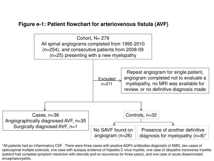
Cohort, N= 279 All spinal angiograms completed from 1995-2010 (n=254), and consecutive patients from 2008-09 (n=25) presenting with a new myelopathy. Figure e-1 : Patient flowchart for arteriovenous fistula (AVF).

E N D
Cohort, N= 279 All spinal angiograms completed from 1995-2010 (n=254), and consecutive patients from 2008-09 (n=25) presenting with a new myelopathy Figure e-1: Patient flowchart for arteriovenous fistula (AVF) Repeat angiogram for single patient, angiogram completed not to evaluate a myelopathy, no MRI was available for review, or no definitive diagnosis made Excluded n=211 Cases, n=36 Angiographically diagnosed AVF, n=35 Surgically diagnosed AVF, n=1 Controls, n=32 No SAVF found on angiogram (n=26) Presence of another definitive diagnosis for myelopathy (n=8)* *All patients had an inflammatory CSF. There were three cases with positive AQP4 antibodies diagnostic of NMO, two cases of opticospinal multiple sclerosis, one case with autopsy evidence of hepatitis C virus myelitis, one case of idiopathic transverse myelitis (patient had complete symptom resolution with steroids and no recurrence for three years), and one case of acute disseminated encephalomyelitis.