Prostate probe with SPECT technique
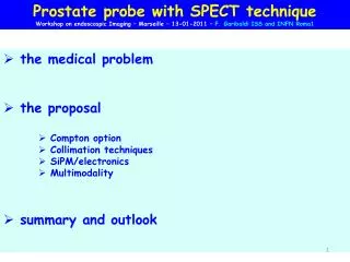
Prostate probe with SPECT technique Workshop on endoscospic Imaging – Marseille – 13-01-2011 – F. Garibaldi ISS and INFN Roma1. the medical problem the proposal Compton option Collimation techniques SiPM/electronics Multimodality summary and outlook.

Prostate probe with SPECT technique
E N D
Presentation Transcript
Prostate probe with SPECT technique Workshop on endoscospic Imaging – Marseille – 13-01-2011 – F. Garibaldi ISS and INFN Roma1 • the medical problem • the proposal • Compton option • Collimation techniques • SiPM/electronics • Multimodality • summary and outlook
Compton Camera Applications to Bio-medical Imaging (Mattinata 5-7 September 2002) Stem cell workshop – Marseille - December 2008 • Topical SymposiumonAdvanced molecular intraoperative probes assisting surgical interventions • TOF PET workshop • Baia delle Zagare September 2009 next ?
- etherogenous, multifocal, biologically not well understood • The most common cancer in men, in western countries (97% of all cancers in men) (EJNM (2008),35:1019-1025) Global incidence of prostate cancer* thesecond leading causeof cancer death <7.4 7.4-13.8 13.8-24.5 24.5-40.7 40.7-124.8 • - Primary or recurrent cancer confined in the organ can be curatively treated • Thus it is important, at primary diagnosis, follow up and recurrence, to obtain accurate assessment of the diesease stage in order to decide the most effective treatment strategy (EJNM (2008),35:1019-1025) *Age-standardised incidence rates per 100,000 GLOBOCAN 2002
SENSITIVITY 83%SPECIFICITY 17% prostate specific, not cancer specific (prostatitis, prostatic iperplasia, prostate cancer) PSA Selective indication : • PSA > 10 ng/ml • cT3 • Gleason score > 7 CT diagnosis is made from tissue obtained in a blind biopsy Need to consider fundamental changes in the approach to diagnosing prostate cancer In the future, multimodality imaging approach tailored to each patient PSADRETRUS biopsy
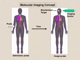
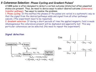
Patient injected with radioactive drug. Drug localizes according to its metabolic properties. Gamma rays, emitted by radioactive decay, that exit the patient are imaged. Radionuclides imaging techniques • Collimator • Only gammas that are perpendicular to imaging plane reach the detector • Readout Electronics • Amplify electrical signal and interface to computer • Scintillator • Converts gammas to visible light • Computer decoding procedure • Elaborate signal and gives image output • Photodetector • Convert light to electrical signal
Compton Camera mechanical collimation PET Multi pinhole
Single photon techniques pros • simple(r) • cheape(r) • extending the radiotracers available • dual tracer looking at two different biological processes cons • - efficiency • spatial resolution
Compton Prostate Imaging Probe Internal Compton Probe External Compton Probe
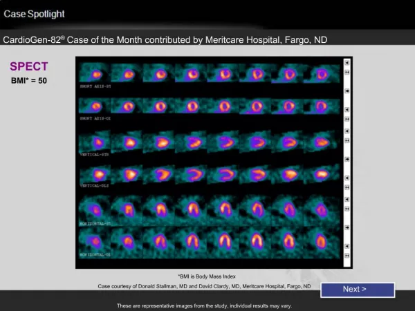
Conventional SPECT Reconstructions 5:1 10:1 15:1 20:1 w / tumor Prostate bkgd 171 and 245 keV, 8.8M events / 40 slices Spatial resolution ~15mm FWHM
Compton Prostate Probe Reconstructions 245 keV only, 1.2 million events, 8mm lesion Prostate 5:1 10:1 15:1 20:1 w / tumor bkgd Spatial resolution ~2mm FWHM
Internal Detector Details 10–12 layers of 1mm thick Si detectors + position and orientation sensor Exploded View Assembled Unit
Detector Packaging Use Tape Automated Bonding (TAB) (Very thin kapton tape with aluminum traces) Kapton microcables “Raw” energy spectrum Detector VATA ASIC Unfolded energy spectrum Kapton “hybrid” board
Demise of the Compton Prostate Probe • Decreasing interest in imaging single photon agents • “Coincidence” PET cameras not reimbursed by HCFA • Technology ultimately was a bit far off
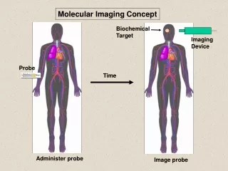
Radionuclides Single photon • 111In-ProstaScint is not a good radiotracer but a new one proposed by M. Pomper looks promising. The single photon endorectal probe provides 2D imaging. We have to try to have 3 D images ( using multipinhole collimation and/or adding up a SPECT tomograph (spatial resolution would be dominated by the small probe (see later, the PET case))
our proposal -insert scintillator pixels into square holes of the collimator better performances (spatial resolution (?) and sensitivity (thicker scintillator)) -using diverging collimator better performances (reducing scan time) -using multipinhole collimation better performances (increasing sensitivity, tomographic recinstruction)
TRANSIZION ZONE Around to prostatic urethra 25% prostatic parenchyma 20% prostate cancer CENTRAL ZONE Encircles the Eyaculatory ducts 10% prostatic parenchyma 1-5% Prostate cancer PERIPHERAL ZONE Region postero-lateral 70% prostatic parenchyma ≥ 70% prostate cancer CANCER LOCALIZATION
Radiotracers issue Radiotracers available for SPECT and PET (from “New agents and Techniques for Imaging prostate cancer” A. Zahreer, S. Y. Cho, M. Pomper”, to be published on JNM) SPECT: Prostascint, Bombesyn,99mTechnetium nanocolloid (limphonodes), other coming soon… PET C—11 Choline, F-18-Choline, Ga-68 Dotabomb (Hofmann (Rome workshop)) many others coming… (collaboration with Johns Hopkins for testing in ISS (mice models for prostate available) and/or at JHU)
PET MRI advantages and issues and fMRI ? • - CITRATE that is present in the normal prostate • CREATINAthat may increase in the phlogosis and all the proliferative processes • - COLINEspecific for a neoplastic transformation “In conclusion, our results confirm that simultaneous PET and high-field-strength MR imaging with LSO-APD–based PET detectors is feasible without sacrificing the quality of images obtained with either system.” M.S. Judenhofer et al. Nature Medicine, Vol. 14 N 4, pg. 439, April 2008
Summary and Conclusions - prostate cancer detection, diagnosis and staging very difficult • standard imaging systems suffer from VERY low specificity - better radiotracers + multimodality can be the solution • single photon techniques are an option because • simpler and cheaper than PET • dual tracer • multimodality with MRI possible • FDA approved for tests on humans • better radiotarcers coming soon - Using also an external SPECT? - Prospectives: Compton