Download
1 / 40
400 likes | 428 Vues
Explore the functions and anatomy of the respiratory system, including ventilation, gas exchange, and defense against pathogens. Discover how the system aids in maintaining homeostasis and enabling vocal communication.

E N D
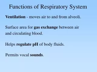
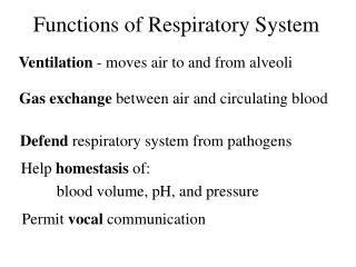
Functions of Respiratory System Ventilation - moves air to and from alveoli Gas exchange between air and circulating blood Defend respiratory system from pathogens Help homestasis of: blood volume, pH, and pressure Permit vocal communication
Respiration • Ventilation- environment and lungs • External Respiration – alveoli and blood • Internal respiration – Blood and cells • Cellular Respiration – Biochemical inside cell
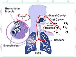
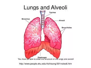
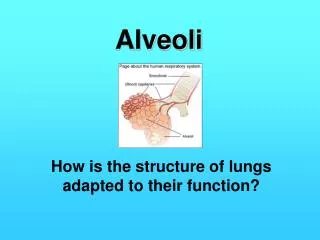
Respiratory organs • Respiratory organs • Nose, nasal cavity, and paranasal sinuses • Pharynx, larynx, and trachea • Bronchi and smaller branches • Lungs and alveoli • Divided into: • upper • lower
Upper respiratory system • Nose • Nasal cavity • Pharynx • Functions • Warm, Filter and Humidify • incoming air • Lower Respiratory System • Larynx • Trachea • Bronchi • Bronchioles • Alveoli
The Nose • Provides an airway for respiration • Moistens and warms air • Filters inhaled air • Houses olfactory receptors
Respiratory Epithelium. • Lines conducting portions • Pseudostratified ciliated columnar epithelium with goblet cells PSCC • Produces mucus to trap foreign particles
cilia rhythmically ‘sweeps’ debris up to be swallowed or expelled at pharynx • Mucus escalator • Alveolar macrophage • Hairs in nose • Cilia - lining respiratory tract
External nares • Open into nasal cavity • Vestibule guarded by hairs • Nasal cavity • Superior, middle and inferior meatuses • Narrow grooves and conchal surfaces • Hard palate • Nasal and oral cavities • Soft palate • Superior nasopharynx and pharynx • Internal nares • Between nasal cavity and nasopharynx
The Pharynx Shared by digestive and respiratory systems • Nasopharynx • Superior portion: from internal nares to uvula. • Oropharynx • Continuous with oral cavity • Laryngopharynx • Between hyoid and entrance to esophagus
Nasopharynx • Superior to the point where food enters • Only an air passageway • Closed off during swallowing • Pharyngeal tonsil (adenoids) • Located on posterior wall • Destroys entering pathogens • Contains the opening to the auditory tube
Oropharynx • Extends from soft palate to the epiglottis • Epithelium is stratified squamous epithelium • Two tonsils in the oropharynx • Palatine tonsils – in the lateral walls • Lingual tonsils – covers the posterior surface of the tongue
Laryngopharynx • Passageway for both food and air • Lined with stratified squamous epithelium • Continuous with the esophagus and larynx
The Lower Respiratory System • Surrounds glottis - air passes through glottis to reach lungs • Epiglottis - prevents solids from entering respiratory system Larynx
During swallowing, elevation of the larynx folds epiglottis over the glottis, steering materials into the esophagus.
Trachea • Submucosa includes “C” rings of cartilage • Tracheal cartilages • Stiffen tracheal walls and protect airway • Posterior wall distorts allowing food passage through esophagus
Left and Right 1o Bronchi • Right and left primary bronchi • Trachea branches within mediastinum • Bronchial tree • Enters lungs at hilus • Root of lung • Bronchus, primary vessels, nerves • Bronchial tree – extensively branching respiratory passageways • Primary bronchi (main bronchi) – largest bronchi • Right main bronchi – wider and shorter than the left
Lungs • Right lung has three lobes • Superior lobar, middle lobar and inferior lobar bronchi • Left lung has two lobes • Superior lobar and inferior lobar bronchi • Cardiac notch
Secondary (lobar) bronchi • Three on the right • Two on the left • Tertiary (segmental) bronchi • Branch into each lung segment • Bronchioles – little bronchi, less than 1 mm in diameter • Terminal bronchioles – less than 0.5 mm in diameter
For clarity, the degree of branching has been reduced: an airway branches approximately 23 times before reaching the level of a lobule.
Fig 24.11
Alveolus – cells and composition • Simple squamous epithelium (type I) • Septal cells (type II) • Produce surfactant • Alveolar macrophages (dust cells) • Patrol epithelium • Engulf foreign particles
Basic structure of a lobule, cut to reveal the arrangement between the alveolar ducts and alveoli.
Respiratory Membrane • Respiratory Membrane (blood-air Barrier) • “point of gas exchange” • Aveolar Epithelium simple squamous epithelia • Fused basement membrane • Capillary endothelium simple squamous epithelia
Respiratory Epithelium • Air epithelia tissue • Nasal cavity stratified squamous • Nasopharynx PSCC • Oropharynx stratified squamous • Laryngopharynx stratified squamous • Larynx PSCC • Trachea PSCC • Rt./Lt. primary bronchi PSCC • Secondary (lobar) bronchi PSCC • Tertiary (segmental) bronchi PSCC • Terminal bronchioles simple cuboidal • Respiratory bronchioles simple cuboidal • Alveolar duct simple squamous • Alveolar sac simple squamous • Alveolus simple squamous
Ep=pscc Sumb=tunica submucosa Cart=tracheal ring Adv=tunica adventitia
Respiratory Muscles Ventilation - movement of air into and out of lungs.
Eupnea - normal quite breathing at rest. Inspiration: volume of thoracic cavity. Muscle activity required: Diaphragm External Intercostals Sternocleidomastoid
When Forcefully exhaling (hypereupnea): Muscles used: Internal Intercostals Rectus abdominis Transverse abdominis, Internal and External obliques.
Bronchial asthma – a type of allergic inflammation • A hypersensitivity to irritants in the air or to stress • Asthma attacks characterized by: • Contraction of bronchiole smooth muscle • Secretion of mucus in airways • Chronic obstructive pulmonary disease (COPD) • Airflow into and out of the lungs is difficult • Obstructive emphysema • Chronic bronchitis • History of smoking
Cystic fibrosis (CF) – inherited disease • Exocrine gland function is disrupted • Respiratory system affected by: • Oversecretion of viscous mucus • Epistaxis – nosebleed • Epiglottitis – inflammation and swelling of the epiglottis • Dyspnea – difficulty in breathing • Apnea – cessation of breathing