BRANCHIAL APPARATUS
1.02k likes | 2.12k Vues
BRANCHIAL APPARATUS. It consists of four components: Mesodermal branchial arches / pharyngeal arches –(5) 2. Ectodermal branchial cleft/ pharyngeal clefts (4) 3. Endodermal branchial pouches / pharyngeal pouches (4) 4. Pharyngeal membranes (4).

BRANCHIAL APPARATUS
E N D
Presentation Transcript
It consists of four components: • Mesodermalbranchial arches /pharyngeal arches –(5) 2. Ectodermalbranchial cleft/pharyngeal clefts (4) 3. Endodermalbranchial pouches /pharyngeal pouches (4) 4. Pharyngeal membranes (4)
Pharyngeal arches are cylindrical mesodermal bars covered by ectoderm and lined internally by the endoderm • Appears in 4th and 5th week of development • Number-6 pairs of pharyngeal arches of which 5th arch disappears early living no important remnants
Derivatives of the Mesodermal pharyngeal arches • Skeletal elements • Muscular elements • Arteries • Nerves
Mesenchyme of pharyngeal arch: • Original : Paraxial and lateral plate - gives rise to musculature of face and neck • Neural crest cells – source of connective tissue components including bones, cartilages, ligaments in oral and facial region.
Skeletal elements • First arch/mandibular arch: • It is differentiated into dorsal maxillary process and ventral mandibular part. • Maxillary process forms Premaxilla, maxilla,zygomatic bone, Squamous part of temporal bone. • The cartilaginous bar of mandibular part is known as Meckel’s cartilage. • Dorsal part of cartilage is ossified to form malleus and incus.
Succeeding part of the cartilage regresses but its fibrous envelop persists as • - the anterior ligament of the malleus & spheno mandibular ligament. • Fibrous membrane of ventral part of the meckel’s cartilage is ossified to form the body of the mandible
Second arch/hyoid arch: • The cartilaginous part of this arch is called Reichert’s cartilage. • Dorsal part of the cartilage is ossified to form stapes of the middle ear. • Succeeding part of the cartilage forms styloid process of temporal bone and stylohyoid ligament . • Ventral part of the cartilage is converted into lesser cornu and upper part of body of the hyoid bone.
Third arch: Dorsal part disappears ventral part is ossified to form greater cornu and lower part of the body of hyoid bone. Fourth arch: Dorsal part disappears ventral part forms the lamina of the thyroid cartilage. Sixth arch: Dorsal part disappears ventral part forms cricoid and arytenoid cartilages.
Muscular derivatives First arch: Tensor tympani, tensor veli palatini, muscles of mastication, anterior belly of digastric and mylohyoid muscles. Second arch: Stapedius, stylohyoid, muscles of the facial expression, posterior belly of digastric, platysma, auricular muscles, epicranius muscles.
Third arch: Stylopharyngeus muscle Supplied by glossopharyngeal nerve Fourth arch: Cricothyroid muscle by external laryngeal nerve which is a branch of superior laryngeal nerve Sixth arch: All intrinsic muscle of the larynx except cricothyroid are supplied by the recurrent laryngeal nerve.
ANAMOLIES: • First arch syndrome: • Results in various congenital anamolies of eyes, ears , mandible & palate. • Due to insufficient migration of neural crest cells into the first arch during the fourth week of development.
2. Treacher collins syndrome:(Mandibulo-facial dysostosis) It is characterised by malar hypoplasia (under development of zygomatic bone), mandibular hypoplasia, down slanting palpebral fissures, defects in lower eyelid & mal formed external ears. 3. Pierre Robin syndrome: Hypoplasia of the mandible, cleft palate & defect in the eyes and ears.
First arch: Mandibular nerve (post trematic), Chordatympani (pre trematic). • Second arch: Facial nerve • Third arch: Glossopharyngeal nerve • Fourth arch: Superior laryngeal nerve • Sixth arch: Recurrent laryngeal nerve
Summary of the fate of aortic arch arteries • First arch:- Mostly disappears except partly for maxillary artery. • Second arch :- Mostly regresses except dorsal part for the stapedial artery. • Third arch :- Ventral part forms common carotid artery dorsal part forms stem of internal carotid artery . • Fourth arch:- Right side it forms proximal part of right subclavian artery, left side it persists part of arch of aorta. • Fifth arch :-Disappears entirely. • Sixth arch :- -On right side the ventral part persists as right pulmonary artery dorsal part disappears . -On the left side the ventral part forms the left pulmonary artery and the dorsal part persist as ductus arteriosusin fetal life /ligamentum arteriosum after birth.
Facts to remember…….. • All the pharyngeal arches are supplied by one nerveexcept….. - First arch (trigeminal – maxillary and mandibular) • Chorda tympani – branch of facial • Only pharyngeal arch that has two processes – first arch (maxillary and mandibular)
Meckel’s cartilage – first arch • Reichert’s cartilage – 2nd arch • Two most important first arch syndromes - Treacher Collins syndrome - Pierre Robin syndrome
Pharyngeal pouches: • 5 pharyngeal pouches. • 5th pouch is rudimentary. • Ventral part of each pouch is obliterated by developing tongue rudiments. • Dorsal part (expect 1st pouch) divides into ventral & dorsal wings.
First pouch: • Dorsal part extends into adjacent mesenchyma as Tubo tympanic recess. • Medial portion of this recess is narrow and persists. as “AUDITORY TUBE”. • Lateral part dilates to form primitive tympanic cavity • Tympanic cavity gives development to, mastoid antrum , mastoid air cells and mucous layer of tympanic membrane.
The blind distal end of the tubo tympanic recess comes in close contact with bottom of the first brachial cleft separated by a thin layer of mesenchyme. • This area forms into future tympanic membrane which is tridermal in development involving the 3 primitive germinal layers. • The cuticular layer from ectoderm, fibrous layer from the mesoderm, and the mucous layer from the tubo-tympanic recess of endoderm.
Second pouch: • Dorsal wing of the 2nd pouch joins with first pouch and contributes in the formation of the tubotympanic recess. • Endodermal cells of the ventral wing of the 2nd pouch proliferate into number of tiny solid buds which extend into adjoining mesenchyme. • Central cells of these buds undergoes destruction leading into formation of tonsillar pits and crypts. • Lymphocytes are derived from the circulating blood or lymph stream.
Due to continuous accumulation of the lymphatic follicles produces inward bulging of the palatine tonsil into the pharynx. • Thus most of the ventral wing of the 2nd pouch is obliterated except a part which persists as the intratonsillar cleft.
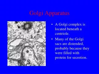
Third pouch: • The dorsal wing of the 3rd pouch is differentiated earlier into inferior parathyroid. • The ventral wing of the 3rd pouch grows caudally as solid thymic rudiment. • Both the wings of pouch communicate with the primitive pharynx by superior pharyngobranchial duct. • The descent of heart and aortic sac allows caudal migration of the thymic rudiment dragging the inferior parathyroid.
As a result inferior parathyroid sweep along the dorsal surface of the lateral lobes of the thyroid gland. • Finally they are disconnected from thymus and gain permanent attachment to the lower pole of the thyroid lobes. • Lymphocytes in the thymic rudiments are from stem cells of the bone marrow.
Fourth pouch: • The dorsal wind of the fourth pouch develops into promodium of the superior parathyroid. • The ventral wing of the 4th pouch joins with rudimentary fifth pouch and forms the caudal pharyngeal complex. • Dorsal wing and complex communicate with the pharynx by the inferior pharyngobrachial duct which eventually rupture.
This complex exhibits three elements: Thymic Element: Incorporated in the development of thymus. Lateral Thyroid Element: Thyroid element of 4th pouch fuses with the median thyroid rudiment. Ultimobranchial body: It plungs into the substance of thyroid rudiment and persists as the parafollicular cells.
Floor of the Primitive Pharynx(Ventral derivatives): • The floor o the primitive pharynx exhibits 3 features. • Tongue • Thyroid Gland • Laryngo-tracheal groove
THE TONGUE: • The tongue appears in the embryo of approximately 4 weeks in the form of two lateral lingual swelling, one medial swelling the tubuerculum impar originates from the first pharyngeal arch. • The 2nd median swelling called the Hypobranchial eminence is formed by mesoderm of 2nd,3rd and part of the 4th arch.
Mucous membrane • Anterior 2/3rd – pair of lingual swellings (endoderm of the first arch) and median tuberculum impar • Posterior 1/3 rd – Hypobranchial eminence (fusion of 2nd, 3rd and 4th arch) • Posteriormost part – Fourth arch • Muscles of the tongue – Occipital myotomes • Connective tissue – Local mesodem
Finally 3rd median swelling formed from the posterior part of the 4th arch gives the development of epiglottis. • Immediately behind the swelling is the laryngeal orifice, which is flanked by the arytenoid swellings. • The epiglottis and the extreme posterior part of the tongue are innervated by the superior laryngeal nerve, reflecting there development from the 4th arch.
Developmental anamolies of the tongue:- Aglossia:- This is due to complete agenesis of the tongue rudiments. Hemi-glossia:- This is caused by the supression of one of the lingual swellings. Bifid tongue:- The anterior part of the tongue splits into two, and is caused by the failure of fusion of the two lingual swellings.
Ankylo-glossia (or) tongue tie: When alveolo-lingual sulcus separates the tongue imperfectly, the movements of the tongue are restricted so much that the lingual speech is disturbed. It is manifested by the shortening of the frenulum linguae.
Development of the thyroid gland • Endodermal diverticulum – thyroglossal duct • Which grows caudally from the floor of the primitive pharynx,behind the tuberculum impar • The lower bifid end proliferates to give thyroid gland • Part of the lateral lobe develops from fourth pharyngeal pouch • Foetal functioning of thyroid begins between 18th & 22nd week
Developmental anamolies of the thyroid: • Thyroglossal cyst (or) fistula: • Lingual thyroid: Arrested caudal growth of the thyroglossal duct may lead to the development of the thyroid gland within the tongue.
Accessory thyroid:- Sometimes nodules of thyroid tissue are found in close proximity to the main gland. Ectopic thyroid:- On rare occasions the thyroid grows in the posterior triangle of the neck/in the thorax. Agenesis of the thyroid:- Complete absence of the thyroid gland is a rare phenomenon. It probably occurs when the anti thyroid antibodies appear within the mother, which might prevent the growth of the foetal thyroid tissue after passing through the placental barrier.
Ectodermal derivatives or clefts: • The ectoderm covering the 1st arch forms: • 1. Skin over the lower jaw • 2. Skin over the upper jaw • 3. Tragus of the auricle • 4. Ectoderm covering the maxillary & mandibular process surrounds the oval fissure and enters into the formation of upper and lower lips, and alveolo labial sulcus. • Early human embryos exhibit usually four ectodermal brachial clefts. • Later only the dorsal part of the first cleft persists and the remaining cleft disappears.
The ventral part of first cleft is obliterated where as dorsal part projects towards the tubotympanic recess of the first pharyngeal pouch results in the formation of external acoustic meatus and cuticular layer of tympanic membrane. • The remaining clefts starts disappearing, before regression epibranchial placodes appear ats at the dorsal end of 2nd, 3rd & 4th clefts by the proliferation of ectodermal cells. • The placodes gradually burrow into the underlying mesenchyme an placodal vesicles which eventually gets disconnected from surface ectoderm and gives rise to development of ganglia of facial, glossopharyngeal and vagus nerves.
The second arch grows much faster than the succeeding arches and come to overhanging them. • The space between the overhanging 2nd arch & 3rd,4th,6th arch is called cervial sinus. • Active proliferation of mesenchymal tissue in 2nd arch causes it to overlap the 3rd and 4th arches and finally it merges with the epicardial ridge in the lower part. • The 2nd,3rd and 4th clefts loose contact with the outside. • Subsequently placodal vesicles degenerate and disappear.
The closure of cervical sinus occurs due to caudal growth of 2nd brachial arch which eventually meets and fuses with epipericardial ridge and the smooth concavity of the side of the neck is restored.
Anamolies: Branchial Cyst: It is painless cystic swelling situated anywhere beneath the sternomastoid muscle, but is more frequently found close to the angle of the mandible. It may of two types: (a) lined by stratified squamous epithelium: A squamous-celled cyst may occur due to failre of obliterationo f the cervical sinus after its initial seperation from the surface epithelium, or due to the persistence of the placodal vesicle. (b) lined by simple columnar epithelium: It may exist when some of the endodermal pharyngeal pouches fail to regress properly.