Download
1 / 31
310 likes | 430 Vues
Explore the essentials of Cardiovascular Physiology, including hemodynamics and determinants of perfusion for optimal tissue oxygenation. Learn about cardiac output, blood pressure, electrical and mechanical functions, and abnormal heart rhythms.

E N D
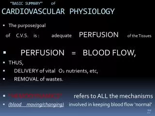
“BASIC SUMMARY” of CARDIOVASCULAR PHYSIOLOGY The purpose/goal of C.V.S. is : adequate PERFUSION of the Tissues PERFUSION = BLOOD FLOW, THUS, DELIVERY of vital O2 nutrients, etc, REMOVAL of wastes. “HEMODYNAMICS” refers to ALL the mechanisms (blood moving/changing) involved in keeping blood flow ‘normal’
DETERMINANTS OF PERFUSION: • 1) CARDIAC OUTPUT Proper Electrical and Mechanical Function of the HEART as a PUMP • BLOOD VOLUME the vascular ‘tank’ must be adequately filled with fluid to supply the need 3) BLOOD PRESSURE : “ FLOW” of a fluid is dependent upon PRESSURE - and is always from “high” pressure area to lower pressure area.
At rest, typically, Cardiac Output is 5 liters per minute(recall that an average man’s total Blood Volume is ~ 5 liters)
CARDIAC OUTPUT depends on 2 ‘functions’ of the heart: • A) ELECTRICAL function (Conduction system) Generation and Propagation of a coordinated Impulse to contract, from atrium to ventricle Conduction system: SA node, AV node, AV Bundle, R and L Bundle Branches, Purkinje system (graphic measure of conduction: electrocardiogram) ECG = “EKG” p wave, QRS complex, t wave B) MECHANICAL FUNCTION 1. CONTRACTILITY of the Myocardium ( strength / force of the contractions) 2. VALVULAR function
Cardiac conduction system: SA node AV node Bundle of His Right Bundle Branch Left Bundle Branch Purkinje Fibers AV Bundle, (Bundle of His)
ELECTRICAL FUNCTION REGULATED RATE AND RHYTHM: Rate -- optimal rate depends on demand. normal at rest 60-100 bpm (Lance Armstrong’s is in the 30’s ) abnormal rates: bradycardia too slow tachycardia too fast RHYTHM -- SHOULD BE REGULAR not skippy or chaotic
Normal sinus rhythm, rate ~72 bpm Nor Onset, supraventricular tachycardia, rate ~120 Normal sinus, rate ~64 bpm Rhythm strips Atrial fibrillation, V.rate varies, ~130-150 Ventricular fibrillation, a terminal rhythm
MECHANICAL FUNCTION CONTRACTILITY of the MYOCARDIUM: the strength / force & completeness of the Contractions (effects of ischemia / necrosis; CAD, HTN drugs, etc) The essence of “Congestive Heart Failure” is usually that of INADEQUATE squeeze capacity of the heart muscle
Cardiac Cycle DIASTOLE - atria contract, Vent. relax; blood flows thru the AV valves, fills Ventricles. (P wave on the EKG) SYSTOLE –Ventricles contract, Blood EJECTED into Aorta and Pulm. Trunk, Art. (QRS complex on the EKG)
VALVULAR FUNCTION • NARROWED VALVE: STENOSIS diminished outflow • INCOMPETENT VALVE: REGURGITATION or INSUFFICIENCY: abnormal “Backflow “ the “ Heart Sounds “ are made by the valves closing – ‘ Lub Dupp lub dupp lub dupp Murmur : swishing sound , made by TURBULENCE of flow can be either from Stenosis or Regurgitation *** abnormal valvular function affects Cardiac Output directly, and indirectly by eventually affecting Contractility of the muscle
CARDIAC OUTPUT • CARDIAC OUTPUT - HOW MUCH BLOOD CAN BE PUMPED each minute? • normal, resting ~ 5 liters / min (roughly the entire blood volume makes one cycle in one minute) • CO = HR x SV CARDIAC HEART RATE X STROKE VOLUME OUTPUT pulse amt blood ejected beats per minute each beat
BLOOD VOLUME Euvolemiahypovolemia volume overload Multiple variants: “Hydration” status (intake -- outgo) diuresis, diarrhea, vomiting, sweating, hyperthermia/ fever, Proper blood production by bone marrow Lack of ‘hemorrhage’, or blood loss complex regulatory mechanisms involving kidney function, endocrine regulatory centers, plasma protein conc., & others
BLOOD PRESSURE • 1. It takes adequate BLOOD Volume for normal BP • 2. It takes adequate ‘FORCE’ on the FLUID • to make it FLOW: • A. Contractions of the Heart generate initial • PRESSURE SURGE, but, the chief determinant of Blood pressure is: • B. VASCULAR RESISTANCE- affects the BLOOD VOLUME IN THE ARTERIES: • Increased resistance VASOCONSTRICTION ^’s BP • (but less flow) • Decreased resistance decr’s BP: VASODILATION • (more flow)
REGULATION of Arterial Resistance • The regulation / changes made in Arterial resistance • in the various ‘REGIONs’ of the body Account for the Alterations /adjustments in • BLOOD FLOW to those areas (increased flow to MUSCLES during exercise, • Incr. Flow to Digestive tract after meals, • ^ flowed to skin to dissipate heat, Whereas Blood Flow to BRAIN and KIDNEYS must remain rel. CONSTANT
Measuring blood pressure: SPHYGMOMANOMETER AND STETHOSCOPE SYSTOLIC BLOOD PRESSURE / DIASTOLIC BLOOD PRESSURE (Korotkoff sounds)
other important effects (on CO, and PERFUSION) • VENOUS RETURN: • THE FLOW OF BLOOD BACK TO THE HEART AFTER • DELIVERY TO THE CAPILLARIES • VEINS: • larger diameter, with elastic walls, increased • CAPACITANCE compared to corresp. Artery • IMPORTANT - GRAVITY generally impedes VENOUS return ----- so, • VEINS have series of one way VALVES - which keep • the blood from flowing backwards
Also: There are 2 FUNCTIONAL VENOUS ‘PUMPS’: 1. SKELETAL MUSCLE CONTRACTIONS, and 2. RESPIRATORY MOTIONS create negative inspiratory pressure, which ‘’SUCKS’ VENOUS BLOOD INTO THE THORAX, TOWARD THE HEART
Modifications that promote venous return: Large lumens Valves • Muscular pump – skeletal muscle activity “milks” blood toward heart • Respiratory pump – pressure changes during breathing move blood toward heart
And FINALLY: CAPILLARIES must mention : CAPILLARIES – Where the ‘ACTION IS’ regarding EXCHANGE of substances between the Vascular & INTERSTITIAL spaces , at the Cells / Tissues level. (fluid, o2, co2 WBC’s, molecules, etc ) can diffuse back and forth ( importance of hydrostatic and osmotic pressure )
Structure of Capillaries: ONLY 1 layer of endothelium and A basement membrane, with “SPACES” for DIFFUSION - no muscle or connective tissue covering. A large number of capillaries in a specific region is called a CAPILLARY BED
5. Circulatory Shock (or circulatory failure, a state of hypoperfusion) Inadequate blood flow to meet cellular needs. Hypovolemic shock – due to decreased blood volume. Septic shock (or vascular shock) – due to inappropriate vasodilation, brought about by response to overwhelming infection. Cardiogenic shock – due to poor heart function.