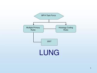
Lung Nodules
Lung Nodules. Frans Naudé. Definition of Pulmonary nodule. Rounded opacity , moderately well defined < 3cm in diameter. Web p 97. General Approach to lung nodules. Position Is it a lung nodule? Skin tags, nipple shadows, bone lesions Distribution in the lung

Lung Nodules
E N D
Presentation Transcript
Lung Nodules Frans Naudé
Definition of Pulmonary nodule • Rounded opacity , moderately well defined • < 3cm in diameter Web p 97
General Approach to lung nodules Position • Is it a lung nodule? • Skin tags, nipple shadows, bone lesions • Distribution in the lung Number of Nodules ( SPN, Multiple) Compare with previous radiographs Interpreting CXR p102
Lung nodules: Imaging Modalities • CXR • CT ( HRES) • PET/CT : F18-FDG
Description of pulmonary nodule • Pattern of distribution (Relationship to fissures, pleura, secondary lobules) • Edge characteristics (sharp, poorly circumscribed, ground glass) • Morphology ( branching/ tree in bud) • Size: • Pulmonary nodule <3cm • Small nodule < 1cm Web p97 High resolution CT of the lung
Secondary pulmonary lobule interlobular septa centrilobular region lobular lung parenchyma Blue = Pulmonary veins Green = lymphatic's Yellow= bronchiolar branches Red = Arteries White = Connective tissue Computed Tomography of the lung p9
Secondary pulmonary lobule Prof Naidich
HRES CT • Interstitial nodules vs Air space nodules
WEB-Algorithmic approach to nodules P120 High resolution CT lung ,Web - approach
HRES CT High resolution CT lung ,Web – Algorithm 4
Airspace nodules • = centrilobular distribution • = no pleural/septal nodules • Ground-glass opacification/ less dense than adjacent blood vessels
Tree – in - Bud • PT with TB • Indicative of endobronchial spread P83, Computed Tomography of the Lung,Verschakelen
Infective bronchiolitis • Tree in Bud appearance • Bronchial wall thickening Computed Tomography of the Lung,Verschakelen
HRES CTTree in bud absent High resolution CT lung ,Web - approach
Poorly defined hazy ground glass nodules • Respiratory bronchiolitis • Langercellhistiocytosis • Lymphocytic interstitial pneumonitis
Interstitial nodules • = pleural/ septal predominance
HRES CTPerilymphatic High resolution CT lung ,Web - approach
Perilymphatic disease • Clustered nodules • Adjacent to fissures and pleural surfaces and along central vascular structures • DDX: Sarcoid, silicosis, CWP. • Rare: Amyloid ,LIP
Sarcoidosis P83, Computed Tomography of the Lung,Verschakelen
Silicosis Web p305
Coal workers pneumoconiosis Web p 306 Diffuse pattern more in favour of CWP or silicosis than sarcoidosis
HRES CTRandom High resolution CT lung ,Web - approach
Random nodules • Sharply define,+- feeding vessel DDX • Metastases: lung, breast, kidney, colon, melanoma, thyroid , pancreas • Infection: Milliary TB, septic emboli, fungal infection • Vasculitis • Langercellhistiocytosis
Metastases • Random • Basilar predominance P82, Computed Tomography of the Lung,Verschakelen
Perilymphatic vs. centrilobular TB Centrilobular changes : nodules, tree-in-bud, branching lines Sarcoidosis - Fissural and subpleural nodules
Perilymphaticnodules. Nodules are immediately in contact with interlobular septa and the visceral pleura (B) Centrilobular nodules. Nodules are positioned 5 - 10 mm from costal and visceral pleural surfaces and interlobular septa.
References • High resolution CT of the lung, Web, Naidich • CT of the lung, Verschakelen, De Wever • Prof Naidich RSSA lecture • High-Resolution CT of the Lung: Patterns of Disease and Differential Diagnoses, RadiolClin N Am 43 (2005) 513 – 542 • Imaging of Interstitial Lung Disease, RadiolClin N Am 43 (2005) 589 – 599
SPN Def: focal area of increased round /oval density in the lung parenchyma measuring less than 3cm, Cause : infection, malignancy , inflammation, vascular, congenital Risk : 30-40% malignant
Approach to SPN • Morphology: • - Size ( smaller more likely benign) • - margins and contours
Internal characteristics • Homogeneous attenuation (55% benign, 20%malignant) • Pseudocavitation and air bronchograms: lymphoma or bronchioalveolar cancer • Benign cavitation : smooth ,thin walls (<4mm) • Malignant cavitation: thick irregular walls( >16mm) • Intranodular fat = hamartoma • Benign calcification : • post infection: central, diffuse solid, laminated , • hamartoma : popcorn like • Malignant calcification: diffuse,amorphous,punctate • Metastatic osteosarcoma: high attenuation nodule
25-39% malignant nodules classified as benign on radiological morphology assessment • growth rate assessment: doubling rate ( increase in diameter of >26%) for malignant nodules between 30-400 days • Clinical data: age, risk factors, previous malignancy
Distribution of lung nodules • Cancer – basal predominance • Breast CA, Colon, Renal often metastasize to lung Interpreting CXR p100
Size of lung nodules • Mayo clinic CT screening trial • ( in patients with no history of cancer) • <3mm = less than 0,2% malignant • <5mm = fewer than 1% malignant • 4-7mm = 0,9% malignant • 8-20mm = 18% malignant • >20mm = 50% malignant Radiology Nov 2005 p 397
Follow-up National Lung Screening Trial • nodules smaller than 4mm • return for screening after 12 months, without interval scans or other work-up Radiology Nov 2005 p 397