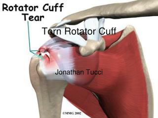
Rotator Cuff Disorders
Rotator Cuff Disorders. Glenohumeral Joint. Shallow (“golf ball sitting on a tee”) Inherently unstable (maximizes ROM). Radiographic Anatomy. GH Joint Stabilizers. Static stabilizers Glenohumeral ligaments, Glenoid labrum and Capsule Dynamic stabilizers

Rotator Cuff Disorders
E N D
Presentation Transcript
Glenohumeral Joint • Shallow (“golf ball sitting on a tee”) • Inherently unstable (maximizes ROM)
GH Joint Stabilizers • Static stabilizers • Glenohumeralligaments, Glenoidlabrum and Capsule • Dynamic stabilizers • Predominantly Rotator Cuff muscles • Scapular Stabilizers • Trapezius, leavator scapulae, serratus anterior, rhomboids • Contraction of the long head of the biceps tendon • Coordinated scapulothoracic rhythm • Proprioceptive mechanoreceptors in the joint capsule
Static Stabilizers(glenohumeral ligaments, glenoid labrum and capsule)
Glenoid Labrum Fibrous ring attached to the glenoidarticular surface through a fibrocartilagenous transition zone The labrum functions as an anchor point for the GH ligaments and the biceps tendon Deepens the glenoid socket and enhances stability The superior and antero-superior portions of the labrum,areless vascular than the posterior and inferior parts This decreased vascularity of the superior labrum may explain the vulnerability of this area to disruption
GH Ligaments • Superior GHL • Stabilizer of the adductedshoulder. • limits posterior translation with the arm in forward flexion, adduction, and internal rotation, • Prevents anterosuperior migration of the humeral head • Middle GHL • limit both anterior and posterior translation of the arm at 45 degrees of abduction and 45 degrees of external rotation • provide anterosuperiorstability • Inferior GHL • The primary restraint to anterior, posterior, and inferior GH translation with the arm at 45 to 90 degrees of abduction and external rotation
Static Stabilizers(glenohumeral ligaments, glenoid labrum and capsule)
Dynamic Stabilizers • The RTC muscles as well as the scapular rotators contribute to stabilization by enhancing the concavity–compression mechanism. • Contraction of the long head of the biceps tendon • Coordinated scapulothoracicrhythm • Proprioceptivemechanoreceptors in the joint capsule
The Rotator Cuff Lateral portions of Infraspinatus, Supraspinatus, Teres minor and Subscapularis muscles and their conjoint tendon The main function of the conjoint structure is to draw the head of the humerus firmly into the glenoid socket and stabilize it there when the deltoid muscle contracts and abducts the arm The musculotendinous cuff passes beneath the coracoacromial arch, from which it is separated by the subacromial bursa During abduction of the arm the cuff slides outwards under the arch The deep surface of the cuff is intimately related to the joint capsule and the tendon of the long head of the biceps
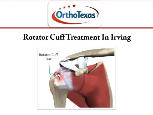
Rotator Cuff Disorders • Supraspinatusimpingement syndrome and tendinitis • Tears of the rotator cuff • Acute calcifictendinitis • Biceps tendinitis and/or rupture
Rotator cuff pain typically appears over the front and lateral aspect of the shoulder during activities with the arm abducted and internally rotated • It may be present even with the arm at rest • Tenderness is felt at the anterior edge of the acromion • Pain and tenderness directly in front along the delto-pectoral boundary could be associated with the biceps tendon • Localized pain over the top of the shoulder is more likely to be due to acromioclavicular pathology • Pain at the back along the scapular border may come from the cervical spine
Supraspinatus Impingement Syndrome and Tendinitis (leading to cuff tear) • Painful disorder arises from repetitive compression or rubbing of the tendons (mainly supraspinatus) under the coracoacromialarch • In the normal shoulder, the coordinated muscle tension within the rotator cuff compresses the humeral head, keeping it centered within the glenoidfossa • Any process that interferes with the rotator cuff's capability to keep the humeral head centred or that compromises the normal coracoacromial arch, including calcium deposits, thickened bursae, and an unfusedosacromiale, can lead to impingement of the rotator cuff
In 1986, Bigliani and Morrison described three variations of acromial morphology. • Type I is flat • type II curved and type III the hooked acromion • They suggested that the type III variety was most frequently associated with impingement and rotator cuff
The impingement process has three chronologic stages: • Stage 1 (Sub-Acute Tendonitis, Painful Arc Syndrome) • Stage 2 (Chronic Tendonitis / Partial Thickness Tear • Stage 3 (Rotator Cuff Disruption / Full Thickness Tear)
Stage 1 (Sub-Acute Tendonitis, Painful Arc Syndrome) • Acute bursitis with subacromialedema and hemorrhage • As the irritation continues, the bursa loses its capability to lubricate and protect the underlying cuff and tendonitis of the rotator cuff develops
Patient Presentation • Insidious pain develops over a period of weeks to months • The patient's history will usually consist of pain with overhead activity, reaching, lifting, and throwing. • They may have a job or recreational activity that involves repetitive overhead movement (painting, tennis) • A long day of overhead activity may increase symptoms to the point where the patient seeks medical attention • The pain usually occurs over the anterolateralaspect of the shoulder, and the patient may point to this specific area • It may radiate down to the deltoid insertion • Very often the patient may report pain at night, exacerbated by lying on the involved shoulder or sleeping with the arm overhead.
Physical Exam • Careful evaluation of the cervical spine to rule out a neurologic problem such as a herniated cervical disc that can mimic shoulder pathology. This is especially true if a patient presents with bilateral symptoms • Feel For Supraspinatous tenderness • Point tenderness is most easily elicited by palpating this spot with the arm held in extension, thus placing the supraspinatus tendon in an exposed position anterior to the acromionprocess • With the arm held in flexion the tenderness disappears
Impingment Tests 1- Painful Arc test 2- Neer Impingement Sign 3- Hawkin’s Impingement Sign Individually, neer and hawkins tests have been shown to be sensitive but not very specific for diagnosing impingement. When combined, these two tests have a negative predictive value greater than 90%
Painful Arc Test • On active abduction scapulohumeralrhythm is disturbed and pain is aggravated as the arm traverses an arc between 60 and 120 degrees. • Repeating the movement with the arm in full external rotation may be much easier for the patient and relatively painless
Neer sign: stabilize the patient's scapula and internally rotate while raising the arm passively in forward flexion • This decreases room available in the subacromial space, thus causing the rotator cuff and overlying bursae to be compressed under the coracoacromial arch.
Hawkins sign the patient's arm is passively flexed to 90 degrees. • The elbow is also bent to 90 degrees and the arm is forcibly internally rotated • This brings the greater tuberosity under the acromion, compressing the cuff and bursae
Stage 2 (Chronic Tendonitis / Partial Thickness Tear • Subacute tendinitis is often reversible, settling down gradually once the initiating activity is avoided • If ignored, inflammation and possible partial thickness tears of the rotator cuff
The patient, usually aged between 40 and 50 • History of recurrent attacks of subacute tendinitis • The pain settling down with rest or anti-inflammatory treatment, only to recur when more demanding activities are resumed • Pain is worse at night • The patient cannot lie on the affected side and often finds it more comfortable to sit up out of bed. • Pain and slight stiffness of the shoulder may restrict even simple activities such as hair grooming or dressing. • Impingement, Neer and Hawkins signs are positive • In addition there may be signs of bicipital tendinitis: tenderness along the bicipital groove and crepitus on moving the biceps tendon • Small, unsuspected tears are quite often found during arthroscopy or operation
Stage 3 (Rotator Cuff Disruption / Full Thickness Tear) • As the process continues the wear on the tendon results in a full thickness tear stage III • The tendon of the long head of biceps, lying adjacent to the supraspinatus, also may be involved and is torn
Large tears of the cuff eventually lead to serious disturbance of shoulder mechanics • The humeral head migrates upwards, abutting against the acromionprocess, and passive abduction is severely restricted • Abnormal movement predisposes to osteoarthritis of the gleno-humeral joint Occasionally • This progresses to a rapidly destructive arthropathyMilwaukee shoulder(named after the city where it was first described
A full thickness tear may follow a long period of chronic tendinitis, but occasionally it occurs spontaneously after a sprain or jerking injury of the shoulder • There is sudden pain and the patient is unable to abduct the arm. • Passive abduction also may, in the early stages, be limited or prevented by pain • If the diagnosis is in doubt, pain can be eliminated by injecting a local anaesthetic into the subacromial space. • If active abduction is now possible the tear must be only partial • If active abduction remains impossible, then a complete tear is likely.
Imaging For Impingement • X-rays are usually normal in the early stages of the cuff dysfunction, but with chronic tendinitis there may be erosion, sclerosis or cyst formation at the site of cuff insertion on the greater tuberosity • In chronic cases thinning of the acromion process and upward displacement of the humeral head. • Osteoarthritis of the acromioclavicular joint is common • In older patients and in late cases the glenohumeral joint also may show features of osteoarthritis.
Magnetic resonance imaging MRI effectively demonstrates the structures around the shoulder and gives valuable information (regarding lesions of the glenoidlabrum, joint capsule or surrounding muscle or bone). • However, it should be remembered that up to a third of asymptomatic individuals have abnormalities of the rotator cuff on MRI • Changes on MRI need to be correlated with the clinical examination
Ultrasonography has comparable accuracy with MRI for identifying and measuring the size of full thickness and partial thickness rotator cuff tears • It has the disadvantage that it cannot identify the quality of the remaining muscle as well as MRI • And cannot always be accurate in predicting the reparability of the tendons.
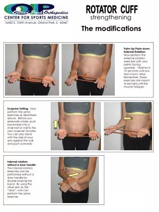
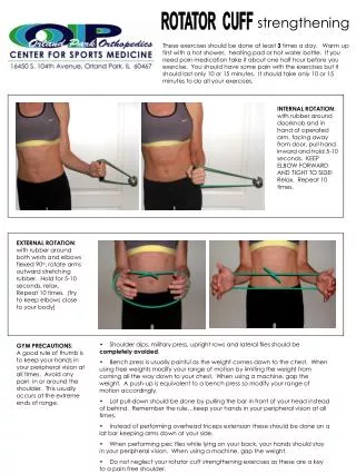
Treatment of Supraspinatus Impingement • Uncomplicated impingement syndrome (or tendinitis) is often self-limiting and symptoms settle down once the aggravating activity is eliminated • The majority of patients with impingement syndrome can be managed conservatively. • The treatment program consists of physiotherapy, activity modification, NSAID, and steroid injections into the subacromial space. • The majority of patients should have a satisfactory result and not require surgery. • The physical therapy program includes soft tissue stretching and strengthening of the humeral head depressors. These are the internal and external rotators. • Strengthening these muscles helps to depress the humeral head and decrease impingement
As range of motion increases, pain levels should decrease. • The scapular stabilizers should also be strengthened. These include the upper and lower trapezius, serratus anterior, and rhomboids. • These muscles contribute to optimal positioning of the scapula during overhead activities. If these muscles fatigue, the scapula is no longer able to keep up with the humerus . The humeral head continues to translate anteriorly and superiorly worsening impingement symptoms. • Strengthening of the deltoid muscle is counterproductive as its action promotes elevation of the humeral head. • Patients with a Type I acromion had a 91% successful result. Patients with Types II and III had less success with 68% and 64%, respectively.
Surgical treatment • The indications for surgical treatment are essentially clinical; • The presence of a cuff tear does not necessarily call for an operation. • Provided the patient has a useful range of movement, adequate strength and well-controlled pain, non-operative measures are adequate.
Indications for surgery • If symptoms do not subside after 3 months of conservative treatment, or if they recur persistently after each period of treatment • Younger patients • Large rotator cuff tears • The operation is subacromial decompression which consists of: • excising the coracoacromial ligament, • undercutting the anterior part of the acromion process • reducing any bony excrescences at the acromioclavicular joint • Repairing rotator cuff tear if present • This can be achieved by open surgery or arthroscopically
Postoperative Management • Pendulum exercises are started within 2 days after surgery. • This is followed by passive range of motion and active-assisted motion. • Full active range of motion can usually be achieved by 3 to 4 weeks. • Light weights and strengthening can begin at 6 weeks. • The overhead athlete should avoid these sports until at least 3 months. • It takes 6 months for complete recovery
Rotator Cuff Disorders • Supraspinatusimpingement syndrome and tendinitis • Tears of the rotator cuff • Acute calcifictendinitis • Biceps tendinitis and/or rupture
Acute Calcific Tendonitis • Acute shoulder pain may follow deposition of calcium hydroxyapatite crystals, usually in the ‘critical zone’ of the supraspinatus tendon slightly medial to its insertion • Cause is unknown
Clinical Features • Affects 30–50 year-olds. • Aching, sometimes following overuse develops and increases in severity within hours, rising to an agonizing pain • After a few days, pain subsides and the shoulder gradually returns to normal. • During the acute stage the arm is held immobile • The shoulder is usually too tender to permit palpation or movement
Treatment • Conservative first (success in 90%) • NSAID • Subacromial injection of corticosteroids • Physiotherapy • Extracorporeal shockwave therapy • Needle aspiration and irrigation (acute cases)
Surgical Treatment • Severe disabling symptoms which have persisted for more than 6 months and are resistant to conservative treatment • Gleno-humeral arthroscopy • Once the calcium deposit is identified, the capsule is carefully incised from the bursal side with a knife in line with fibre orientation of the tendon • A curette is then used to milk out the toothpaste-like calcium deposit. • A subacromial decompression is also usually performed
Biceps Tendinitis • Primary tendinitis involves inflammation of the tendon within the bicipital groove. • To be considered primary, no other pathological findings (such as impingement, bony abnormalities within the groove, or biceps subluxation) should be present • Secondary tendinitis caused by the same causes of impingement syndrome
Anterior shoulder pain (particularly in the region of the bicipital groove) is the hallmark of biceps tendonitis • With biceps tendinitis the pain is usually described as a chronic aching pain, which is worsened by lifting and overhead activities • The pain frequently radiates distally to approximately the mid arm level but rarely radiates proximally. • Inciting events include repetitive activities involving lifting and overhead activities. • There is such a close association between subacromial impingement and biceps tendonitis that the two conditions have closely overlapping symptoms. • They can be very difficult to distinguish
Physical Findings • The hallmark of biceps tendon related pathology is point tenderness in the bicipital groove • The bicipital groove is three inches below the acromion with the arm in 10 degrees of internal rotation • As the arm is internally and externally rotated, the pain should move with the arm • This is distinct from subacromial bursitis where the pain location remains relatively constant despite the position of the arm
Provocative Tests Speed's test With the elbow in extension, the patient flexes the shoulder against resistance from the examiner. Pain in the bicipital groove isconsidered positive Yergason test —The patient attempts to supinate the wrist against resistance (with the elbow flexed at the side). Pain in the bicipital groove isconsidered positive
Treatment • Rest, ice, and NSAID • As symptoms improve, range of motion exercises and strengthening can be added • Subacromial steroid injections or bicipital sheath steroid injections may also be utilized • If conservative fails, surgery • Debridement of the LHB, • Biceps tenotomy (for elderly) • or biceps tenodesis
Rupture of LHB • The patient is usually aged over 50 • While lifting he or she feels something snap in the shoulder and the upper arm becomes painful and bruised. • Ask the patient to flex the elbow: the detached belly of the biceps forms a prominent lump in the lower part of the arm.