Download
1 / 173
1.75k likes | 2.05k Vues
16. The Gastrointestinal System: Fuel for the Trip. Multimedia Asset Directory. Slide 16 Swallowing and Digestion Animation Slide 40 GERD Video Slide 96 Appendicitis Animation Slide 150 Eating Disorders Video Slide 151 Anorexia Nervosa Video Slide 152 Bulimia Video

E N D
16 The Gastrointestinal System: Fuel for the Trip
Multimedia Asset Directory Slide 16 Swallowing and Digestion Animation Slide 40 GERD Video Slide 96 Appendicitis Animation Slide 150 Eating Disorders Video Slide 151 Anorexia Nervosa Video Slide 152 Bulimia Video Slide 153 Dieticians Video Slide 154 Dental Assisting and Dental Hygiene Video
Introduction • The gastrointestinal system has the following functions: • Take in (ingest) raw materials • Break down (digest) raw materials to usable elements • Physically • Chemically • Absorb elements • Eliminate what isn’t usable
Introduction • These functions are accomplished through an array of amazing main and accessory organs with food traveling through what is, essentially, a tube until waste products are eliminated.
Learning Objectives • Locate and describe the functions of the main organs of the digestive system. • Locate and describe the functions of the accessory organs for digestion. • Differentiate between ingestion and digestion, and between chemical and mechanical processing of food. • Trace the journey of a bolus of food from the mouth to the anus.
Learning Objectives • Discuss the structure of teeth. • Describe the various enzymes and chemicals needed for digestion. • Describe common disorders of the gastrointestinal system.
adventitia (ADD ven TISH ah) alimentary tract (AL ah MEN tar ee) appendicitis (ah PEN dih SIGH tis) appendix (ah PEN dicks) cecum (SEE kum) cementum (see MEN tum) cholecystitis (KOH lee sis TYE tis) cholelithiasis (KOH lee lih THY ah sis) chyle (KILE) Pronunciation Guide Click on the megaphone icon before each item to hear the pronunciation.
chyme (KIME) defecation (def eh CAY shun) duodenum (DOO oh DEE num) emulsification (ee MULL sih fih KAY shun) epiglottis (ep ih GLAH tis) esophagus (eh SOFF ah gus) frenulum (FREN you lum) fundus (FUN dus) gingiva (JIN jih vuh) Pronunciation Guide Click on the megaphone icon before each item to hear the pronunciation.
ilium (ILL ee um) jejenum (jee JOO num) labia (LAY bee ah) mastication (MASS tih CAY shun) mesentery (MEZ in TARE ee) pancreatitis (PAN kree ah TYE tis) peristalsis (pair ih STALL sis) pharynx (FAIR inks) plicae circulares (PLY kay sir cue LAIR es) Pronunciation Guide Click on the megaphone icon before each item to hear the pronunciation.
ptyalin (TYE ah lin) pyloric sphincter (pye LOR ik SFINK ter) pylorus (pye LOR us) rugae (ROO gay) serosa (seh ROSE ah) villi (VILL eye) Pronunciation Guide Click on the megaphone icon before each item to hear the pronunciation.
System Overview • The digestive tract, often called the alimentary tract or canal, is a muscular tube that contains the organs of digestion. • The tube begins with the mouth and ends at the anus. • In between these two points are the pharynx, esophagus, stomach, small intestine, and large intestine. • Accessory organs of digestion include the teeth, salivary glands, liver, pancreas, and gallbladder.
Functions of the Gastrointestinal Tract • Ingestion – food enters the mouth • Mastication (chewing) – mechanically grinding food with the teeth and tongue • Digestion – the chemical act of breaking down food into small molecules
Functions of the Gastrointestinal Tract • Secretion – release of acids, buffers, enzymes, and water to aid in the breakdown of food • Absorption – food passes through lining of digestive tract into blood stream • Excretion or defecation – elimination of waste products
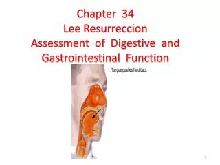
Oral Cavity • The oral cavity is the opening behind the mouth, the hard and soft palate is the roof, the tongue is the floor, and the cheeks are the walls. • The uvula aids in swallowing, directing food toward the pharynx and blocking food from entering your nose. • The mouth receives, tastes, mechanically breaks down, and begins starch digestion.
Click here to view an animation showing Swallowing a Bolus of Food. Back to Directory
Oral Cavity • Tongue • The tongue’s base (area of attachment) and the uvula are the barrier to the next part of the system, the pharynx. • Swallowing • The uvula aids in swallowing, directing food toward the pharynx, and blocking food from entering your nose. • The tongue pushes the food into a ball-like mass, called a bolus, so it may be swallowed – passed to the pharynx.
Oral Cavity • Tongue • Your tongue provides taste stimuli to your brain, determines temperature, and manipulates food. • The lingual frenulum, a membrane under the tongue, keeps you from swallowing your tongue and aids in speaking.
Clinical Application: Sublingual Medication • The area under the tongue has many blood vessels. This sublingual blood vessel network readily absorbs substances and is a rapid means of administering medication. • One medication given by this route is nitroglycerine, used to treat angina. Angina develops as a result of poor oxygen supply to the myocardium because of diminished blood flow.
Clinical Application: Sublingual Medication • Nitroglycerine dilates arteries, improving blood supply and oxygenation – hopefully relieving angina symptoms.
Oral Cavity • Salivary glands (accessory organ) • There are two pairs of salivary glands controlled by the autonomic nervous system. • A large parotid salivary gland is found slightly inferior and anterior to each ear. These are the ones that swell when you get mumps. • The sublingual salivary glands are found under the tongue.
Oral Cavity • Salivary glands (accessory organ) • The submandibular salivary glands are located along both sides of the inner surface of the mandible. • The ducts from these glands empty into the upper portion of the oral cavity.
Oral Cavity • Saliva • The salivary glands produce 1–1.5 liters of saliva daily. • Small amounts of saliva keep the mouth moist, but the idea or presence of food increase production significantly. • Saliva is 99.4% water, and contains antibodies, buffers, ions, waste products, and enzymes. • One enzyme, salivary amylase, speeds the chemical activity of breaking down carbohydrates. • After eating, saliva cleans the oral surfaces, reducing the amount of bacteria that grows in your mouth.
Oral Cavity • Teeth • The first set of teeth you grow as a baby are the deciduous teeth. They will fall out in time. • The 1st tooth appears around 6 months of age. The lower central incisors appear first, with all 20 teeth in place by age 2½. • Between 6 and 12 years these teeth fall out and are replaced by 32 permanent teeth. • Wisdom teeth appear by the time we turn 21.
Oral Cavity • Types of teeth • Incisors are located at the front of the mouth, are blade-shaped, and are used to cut food. • Canine teeth are for holding, tearing, or slashing food. They are also known as eyeteeth or cuspids, and are located next to incisors. • Bicuspids, or premolars, are transitional teeth. • Molars have flattened tops. Both bicuspids and molars are responsible for crushing and grinding food.
Oral Cavity • Tooth anatomy • Teeth have a crown, neck, and root. • The crown is the part you normally see and is covered by the hardest biologically manufactured substance, enamel. • The neck is the transitional section that leads to the root. • Internally, most teeth are made up of dentin, a mineralized bone-like substance. • The next layer is connective tissue, pulp, located in the pulp cavity.
Oral Cavity • Tooth anatomy • The pulp cavity contains blood vessels and nerves providing nutrients and sensation. The nerves and blood vessels get to the pulp cavity via the root canal. • The root is nestled in a bony socket and is held in place by fibers of the periodontal ligament. • In addition, cementum, a soft version of bone, covers the dentin of the root, aiding in securing the periodontal ligament.
Oral Cavity • Tooth anatomy • Healthy gums, or gingiva, help hold the teeth in place. • Epithelial cells form a tight seal around the tooth to prevent bacteria from coming into contact with the tooth’s cementum.
Figure 16-4 (continued) Types, location and, structures of teeth.
Pharynx • There are three parts to the pharynx: • The nasopharynx is primarily part of the respiratory system, blocked by the soft palate. • The oropharynx and the laryngopharynx act as a passageway for food, water, and air. • The epiglottis covers the trachea to prevent food from entering the lungs, forcing food into the opening for the esophagus.
Esophagus • The esophagus is approximately 10 inches long and is connected to the stomach, normally collapsed. • It extends from the pharynx, through the thoracic cavity, through the diaphragm, connecting to the stomach in the peritoneal cavity. • Rhythmic contractions, called peristalsis, push food down the esophagus.
Esophagus • The esophageal walls • Lined with stratified squamous epithelium • Outer layer is adventitia not serosa
Esophageal Sphincters • A muscular ring at the top of the esophagus, called the pharyngoesophageal sphincter, relaxes to open the esophagus so food can enter. • At the entrance to the stomach is the lower esophageal sphincter, or cardiac sphincter, opening the door to the stomach and closing to prevent acidic gastric juices from splashing into the esophagus.
Figure 16-5 The movement of a bolus of food from the mouth to the stomach via the esophagus.
Figure 16-5 (continued) The movement of a bolus of food from the mouth to the stomach via the esophagus.
Click here to view a video on the topic of GERD. Back to Directory
Walls of the Alimentary Canal • The innermost layer, the mucosa, lines the lumen of the canal. • This layer is composed mostly of surface epithelium with some connective tissue and has a thin smooth muscle layer surrounding it. • The mucosa also possesses cells that secrete digestive enzymes to break down foodstuffs and goblet cells that secrete mucus for lubrication.
Walls of the Alimentary Canal • The submucosa is the next layer, and is composed of soft connective tissue. • This layer contains blood vessels, lymph vessels, lymph tissue (called Peyer patches) and nerve endings.
Walls of the Alimentary Canal • The next layer is called the muscularis externa, and is composed of two layers of smooth muscle. • The innermost layer of muscle encircles the canal, while the outer layer of muscle is longitudinal in nature, so it lies in the direction of the canal.
Walls of the Alimentary Canal • The outermost layer, the serosa, is composed of a single, thin layer of flat, serous fluid producing cells supported by connective tissue. • The serosa is the visceral peritoneum in most of the canal.
Figure 16-6 Basic tissue type and structures of the alimentary canal.
Peritoneum • The peritoneum is a serous membrane in the abdominopelvic cavity. • The visceral peritoneum covers the organs, and the parietal peritoneum lines the wall of the abdominopelvic cavity. • Between the layers is a fluid-filled potential space called the peritoneal cavity.
Peritoneum • The peritoneum is a serous membrane in the abdominopelvic cavity. • The peritoneum is different from the other serous membranes. • Some abdominal organs are not surrounded by peritoneum and are called retroperitoneal organs. • The peritoneum has several extensions called mesentery that drape over the abdominal organs.
From the Streets:Swallowed Foreign Bodies • Swallowed foreign bodies are a common reason people seek emergency care. • Objects that lodge in the esophagus usually require intervention. • Treatment may include medications and surgical intervention.
Stomach • The stomach is located in the left side of the abdominal cavity, under the diaphragm, and is covered completely by the liver. • It is approximately 10 inches long with a diameter that depends on how much you just ate. • It can hold up to 4 liters when filled. • Rugae, or folds, help the stomach expand and contract.