Download
1 / 28
310 likes | 619 Vues
Unit 14 The Mouth and Salivary Glands. 2 20 2011 online ed. The Mouth. Also called oral cavity Beginning digestive system Encloses dental arches (rows of teeth) Receives saliva from salivary glands. Anatomy: Mouth. 2 Divisions Oral vestibule: Space between teeth and cheeks

E N D
Unit 14The Mouth and Salivary Glands 2 20 2011 online ed.
The Mouth • Also called oral cavity • Beginning digestive system • Encloses dental arches (rows of teeth) • Receives saliva from salivary glands
Anatomy: Mouth • 2 Divisions • Oral vestibule: Space between teeth and cheeks • Oral cavity or mouth proper • Space between dental arches • Roof formed by hard and soft palates • Floor formed by tongue • Communicates with pharynx posteriorly via oropharynx
Anatomy: Mouth Hard palate Most anterior portion of roof Formed by maxillae and palatine bones Soft palate Begins behind last molar Movable musculomembranous structure
Frenulum • Median vertical band on inferior surface of tongue • Restricts posterior movement of anterior part of tongue
Teeth Purpose? Mastication (chewing)
Tongue Apex- anterior Base- posterior Sublingual space- Part of floor that lies under tongue
Uvula Small pendulous process in center of inferior border of soft palate Purpose? Helps keep food from going wrong way down esophagus when swallowing Singers credit uvula with letting them produce a vibrato, a wavy up-and-down sound.
Tonsils Purpose? Body’s 1st line of defense of immune system They “sample” bacteria and viruses that enter body through mouth or nose at risk of own infection Can be more liability than asset! Common problems: Recurrent bacterial infections (throat or ear) Significant enlargement can obstruct airway causes breathing, swallowing, and sleep problems
Adenoids Like tonsils, adenoids help keep body healthy by: - trapping harmful bacteria and viruses that you breathe in or swallow - containing cells that make antibodies to help body fight infections
Brushing teeth isn’t always enough! • Abscesses, chronic tonsillitis, and infections around tonsils produce foul-smelling, cheese-like formations Pheew! Really bad breathe!
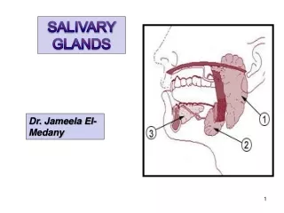
How many salivary glands? • 3 pairs • Parotid (para-otid-situated near the ear) • Submandibular • Sublingual (lingual-of tongue: relating to, using, or similar to) • Produce how much saliva per day? about 1 Liter
What does saliva do? Saliva mixes with food during mastication Softens food Keeps mouth moist Contributes digestive enzymes
Parotid Glands • Largest salivary gland • Lies immediately anterior to external ear • Parotid duct opens into oral vestibule opposite 2nd upper molar
Submandibular Gland • Extends posteriorly from below 1st lower molar to angle of mandible • Greater portion extends below mandible • Submandibular duct opens into mouth on side of frenulum
Sublingual Glands Smallest pair Located in floor of mouth Numerous, small sublingual ducts open into floor of mouth
Sialography • Radiologic exam of salivary glands and ducts using contrast media • Infrequently performed to investigate ducts • CT and MRI have largely replaced this exam
Sialography May be used to demonstrate: • Inflammatory lesions • Tumors • Fistulae (abnormalconnection or passagewaybetween two epithelium lined organs or vessels that normally do not connect) • Diverticula (outpouching of hollow structure inbody) • Strictures (narrowing of bodily passage) • Calculi • Problem -Only one gland can be examined at a time!
Sialography • Submandibular and parotid glands are investigated by this method • Sublingual gland is usually not evaluated this way- Why not? • Difficulty in cannulation (cannula: tube that can be inserted into body, often for delivery or removal of fluid)
Contraindications • Severe infection of gland • Known allergies to contrast media
Procedure Preliminary radiographs Detect conditions that do not require contrast Obtain optimum exposure factors Give pt secretory stimulant 2 to 3 minutes before contrast administration Pt asked to suck on lemon wedge -Opens duct for easy identification
Procedure (cont’d) • Duct is cannulated, not punctured, contrast introduced with fluoroscopic guidance • Take Radiographs • After radiographs, pt sucks on a lemon wedge again to evacuate contrast • Take post-procedure radiographs after 10 minutes to confirm evacuation of contrast
Radiographs Parotid: Tangential Lateral (Right or left)
Tangential parotid • Pt -supine or prone • Parotid area centered to IR • CR perpendicular to IR • Enters lateral surface of mandibular ramus
Parotid Radiographs Same pt with air in cheeks
Lateral Parotid Gland Radiograph Blockage of parotid duct
Axial lateral submandibular gland showing opacification of submandibularduct