Download
1 / 47
480 likes | 530 Vues
Learn about diseases of the salivary glands, including sialolithiasis, tumors, and clinical aspects. Understand common conditions, their manifestations, and treatment options.

E N D
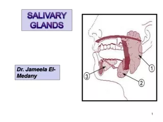
Sialolithiasis &Nonspecific sialadenitis • Submandibular • Stones nidus - impacted food debris - dehydration with decreased secretion • Stone → duct obstruction • Nonspecific interstitial inflammation • Or suppuration (abscess) – Staph. Strep
Sialolithiasis &Nonspecific sialadenitis • Unilateral • Painful – during meals • Purulent ductal discharge + • Recurrent
MUCOCELE • Trauma • Lower lip • Fluctuant swelling • Blockage / rupture of salivary duct → leakage of saliva into the surrounding stroma • RANULA – mucocele of the sublingual
Sjogren’s syndrome • Immune destruction • Salivary – xerostomia (dry mouth) • Lacrimal – keratoconjunctivitis sicca (dry eyes)
TUMOURS • Approx. 30 in number • 2% of all tumours • Benign – 5th – 7th decade • Malignant – later than above
BENIGN TUMOURS • PLEOMORPHIC ADENOMA – 50% • Warthin’s tumour • Oncocytoma • Basal cell adenoma • Canalicular adenoma • Ductal papilloma
PLEOMORPHIC ADENOMA • Benign tumour of glands – ADENOMA • Remarkable histologic diversity - PLEOMORPHIC • Commonest • Also called Mixed tumour – epithelial and connective tissue components
PLEOMORPHIC ADENOMA • Cell of origin → EPITHELIAL epithelial-myoepithelial / ductal reserve cells
PLEOMORPHIC ADENOMAGross • < 6 cms • Bosselated • Well-demarcated / encapsulated / tongue-like protrusions • Cut surface: gray-white / variegated / myxoid / bluish translucent (chondroid)
PLEOMORPHIC ADENOMAMicroscopy • HETEROGENITY ! → PLEOMORPHIC • EPITHELIAL : epithelial & myoepithelial cells arranged in ducts, acini, irregular tubules, strands, sheets NO DYSPLASIA • MESENCHYMAL : myxoid, hyaline, chondroid, osseous
PLEOMORPHIC ADENOMACLINICAL ASPECTS • Slow growing • Painless • Parotid – swelling below & in front of the year • RECURRENT – tongue-like protrusions Enucleation – 25% with Parotidectomy – 4%
CARCINOMA ex PLEOMORHIC ADENOMA OR MALIGNANT MIXED TUMOUR Adenocarcinoma
WARTHIN’S TUMOUR Papillary cystadenoma lymphomatosum Adenolymphoma
WARTHIN’S TUMOUR Gross • Encapsulated • Cut surface : pale-grey narrow cystic / cleft-like / convoluted spaces contain serous / mucinous fluid
WARTHIN’S TUMOUR Microscopy Cystic spaces - Narrow / cleft-like CYSTADENOMA Lining – double-layered epithelium – infoldings PAPILLARY • Inner (lumen) – palisade of columnar cells with abundant finely granular eosinophilic cytoplasm (oncocytic) • Outer (below the first) – cuboidal / polygonal Stroma - dense lymphoid tissue (germinal centres + ) LYMPHOMATOSUM
WARTHIN’S TUMOUR • HISTOGENESIS ? Aberrant inclusions of lymphoid tissue in salivary glands
WARTHIN’S TUMOUR CLINICAL ASPECTS • Benign • Very rarely recurs
MALIGNANT TUMOURS • Mucoepidermoid ca • Acinic cell ca • Adenoid cystic ca • Malignant mixed tumour
Malignant tumours • Old age • Max (70-90%) – sublingual • Malignant tumours more in smaller glands
MUCO-EPIDERMOID CARCINOMA • 15% • 60-70% in the parotid, also in others • MOST COMMON MALIGNANT • Most common radiation-induced
MUCO-EPIDERMOID CARCINOMAGross • < 8 cms. • Apparently circumscribed / infiltrative • Cut surface : pale grey-white small mucin-filled cysts
MUCO-EPIDERMOID CARCINOMAMicroscopy • Mixtures of threecell types arranged in cords, sheets, cystic 1. mucus-secreting cells 2. intermediate cells (hybrid) squamoid features with small to large mucus- filled vacuoles 3. squamous cells
MUCO-EPIDERMOID CARCINOMA • LOW GRADE – Mucus cells predominant • HIGH GRADE – squamous cells predominant
MUCO-EPIDERMOID CARCINOMA CLINICAL COURSE Low gradeHigh grade Spread Local invasion Metastasis(30%) Recurrence 15% 25-30% 5-yr survival >90% 50%
ADENOID CYSTIC CARCINOMA • Mouth – minor, rare in parotid • Nose, paranasal sinuses • Upper airways
ADENOID CYSTIC CARCINOMA Gross • Small • Poorly encapsulated, infiltrative • Gray-pink
ADENOID CYSTIC CARCINOMA Microscopy • Small cells with dark compact nuclei, scanty cytoplasm • Arranged in tubules, solid, cribriform • SPACES BETWEEN CELLS FILLED WITH HYALINE MATERIAL – basement membrane material • Perineural invasion - pain
ADENOID CYSTIC CARCINOMA CLINICAL ASPECTS • Malignant • Slow growing • Stubbornly recurrent • Painful – perineural invasion
Acinic cell carcinoma (tumour) • Most PAROTID – composed of serous cells
Acinic cell carcinoma Gross • Small • Encapsulated
Acinic cell carcinoma Microscopy • Look like serous cells • Clear cytoplasm • Solid sheets, microcystic, glandular, follicular, papillary • Little anaplasia
Acinic cell carcinoma CLINICAL ASPECTS • Good prognosis • Recurrence rare