Download
1 / 26
270 likes | 714 Vues
Patellofemoral Pain. Objectives. Understand the anatomy of the patellofemoral joint Learn 3 causes of PFPS Understand the muscular imbalances that lead to PFPS Understand how the ankle, knee and hip effect patella tracking

E N D
Objectives • Understand the anatomy of the patellofemoral joint • Learn 3 causes of PFPS • Understand the muscular imbalances that lead to PFPS • Understand how the ankle, knee and hip effect patella tracking • Design/ Follow a rehabilitation programto safely return the athlete to sport
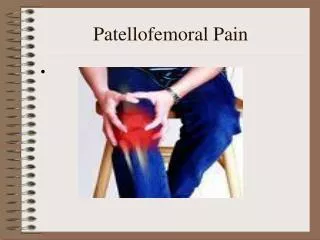
What is Patellofemoral Pain?? • Anterior knee pain resulting from physical and biomechanical changes in the patellofemoral joint • Pain occurs with activity, prolonged sitting and increases with ascending/descending stairs • Classified as overuse or overload injury
The Knee The bony anatomy consists of the distal femur with its two condyles, the proximal tibia with its two tibial plateaus and the sesamoid bone in the quadriceps tendon, the patella.
Patellofemoral Joint • The patella is a sesamoid bone in the quadriceps tendon • It articulates with the intercondylar (trochlear) groove on the anterior aspect of the distal portion of the femur • The patella tendon inserts on the tibia at the tibial tuberosity
The Patella • The patella moves superior, inferior, tilts and rotates as the knee flexes and extends. This is referred to as tracking. • As the knee flexes the patella slides down in the trochlear groove and slides up as the knee extends • The patella also tilts medially and laterally and internally and externally rotates • The posterior surface of the patella contacts the femur at various points and is composed of facets designed to contact at specific points during flexion and extension • Tracking is controlled by muscle contraction to ensure appropriate movement of the patella in the trochlear groove
Patella Tracking • Improper patella tracking is thought to be one of the causes of patella femoral pain syndrome • Maltracking of the patella occurs when the patella no longer remains centered in the trochlear groove, • Abnormal biomechanics results in increased pressure on the patellar joint surface. • Repetitive maltracking causes abnormalities within the articular cartilage, chronic inflammation and pain
Forces effecting patella tracking • Q-angle • Pes Planus (pronation) • Muscular Imbalances
Angle between rectus femoris and patella tendon. Formed by measuring from ASIS to middle of the patella and from mid-patella to tibial tubercle Normal angle is 10* in males, 15-20 in females Increased angle results in lateral patella tracking Causes of increased Q-angle: genu valgus, internal rotation of the femur and a wide pelvis Q angle
“Flat foot” “over-pronation” Causes a compensatory internal rotaion of the tibia or femur Results in increased Q-angle and increased lateral patella tracking Pes Planus
Muscular Imbalances • Vastus medialis obliqus(part of the quadriceps) weakness • Tight Iliotibial band • Tight Hamstrings • Weak Hip abductors • Tight Hip flexors • Tight Plantarflexors
Muscular Imbalances • Weakness of the VMO allows the patella to track too far laterally • A tight iliotibial band places excessive lateral force on the patella and can also externally rotate the tibia, resulting in excessive lateral tracking of the patella • Tight hamstrings place more posterior force on the knee, causing pressure between the patella and femur to increase • Abductor (gluteus medius) strengthening helps to stabilize the pelvis. of the hip external Weak abductors results in compensatory foot pronation; • Tight calves can lead to compensatory foot pronation and, like tight hamstrings, can increase the posterior force on the knee
Recognition • Diffuse anterior knee pain • Edema • Crepitus • + movie –goer’s sign ( pain with prolonged sitting)
Recognition • X-rays can be taken to rule out the presence of a fracture or Osgood-Schlatters • MRI should be taken to rule out a ligament or meniscus injury.
Evaluation It is important to keep in mind there is no traumatic cause! Evaluating for PFPS involves lower extremity screening for the presence of biomechanical deficiences in strength, ROM and patella tracking. The cause of the pain could be from the hip, knee or ankle.
Evaluation Ankle Assessment: Does the athlete excessively pronate? Does the athlete have tight gastrocs?
Evaluation Knee Assessment: Assess patella alignment for a “squinting” position. In standing, the patella will look turned in. “Special Tests” • Clarke sign: The athlete is sitting with knee extended, press down proximal to the top of the patella and ask the athlete to contract the quadriceps as you press down. Positive test= pain in the patella and unable to hold the contraction • Patella Compression Test: The athlete sits with a towel rolled under the knee to position in 20* flexion. Press down on the patella. Positive test = pain in patella.
Evaluation Hip Assessment: • Thomas test: Measures hip flexor flexibility • Ober Test: Measures ITB flexibility
Rehabilitation • Manage pain and inflammation: rest from painful activities, ice • Strengthen weak muscles: • Strengthen to VMO to decrease patella lateral tracking This is achieved through hip adduction exercises, quadriceps isometrics • Strengthen hip musculature to decrease patella lateral tracking This is achieved through glute strengthening exercises 3. Stretch hip flexors, ITB, hamstrings and gastrocs
Rehabilitation Restore the biomechanical abnormalities: Lower extremity strengthening exercises to strengthen the hip and quads include: Step-ups Step-downs Squats Lunges All exercises should be pain free and done with proper form. A sign of weakness is allowing the femurs to internally rotate and knees to come too far forward over the toes.
Rehabilitation • Please see attached word document for exercises
Return to play The athlete should expect approximately 6 weeks of physical therapy. Once the athlete has no knee pain with normal activity, he or she can return to sport.
Prevention • Recognize signs of lower extremity weakness and tightness • Promote calf stretching, hamstring stretching, hip stretching.
References • Tyler Timothy, Stephan Nicholas, Michael Mullaney, Malachy McHugh. " The role of hip muscle function in the treatment of patellofemoral pain syndrome” The American Journal of Sports Medicine. (2006) 34 (4)630-635. • Mark S. Juhn. “Patellofemoral Pain Syndrom: A review and guidelines for treatment” The American Family Physician. (1999) 3. Carrie Halli, Lori Brody (2005).Therapeutic Exercise; moving toward function. New York: Lippincott williams & Wilkins 4. Web site:www.med.umich.edu/1libr/guides/knee/kneeart.htm