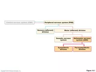
Central Nervous System (CNS)
Central Nervous System (CNS). CNS = Brain + spinal cord Surface anatomy includes cerebral hemispheres , cerebellum , and brain stem. Basic Pattern of the Central Nervous System. Spinal Cord Central cavity surrounded by a gray matter core

Central Nervous System (CNS)
E N D
Presentation Transcript
Central Nervous System (CNS) • CNS = Brain + spinal cord • Surface anatomy includes cerebral hemispheres, cerebellum, and brain stem
Basic Pattern of the Central Nervous System • Spinal Cord • Central cavity surrounded by a gray matter core • External to which is white matter composed of myelinated fiber tracts • Brain • Similar to spinal cord but with additional areas of gray matter • Cerebellum has gray matter in nuclei and cortex • Cerebrum has nuclei and additional gray matter in the cortex
Basic Pattern of the Central Nervous System Figure 12.4
Ventricles of the Brain Figure 12.5
Ventricles of the Brain • Arise from expansion of the lumen of the neural tube • The ventricles are: • The paired C-shaped lateral ventricles • The third ventricle found in the diencephalon • The fourth ventricle, dorsal to the pons
Cerebral Hemispheres • Form the superior part of the brain and make up 83% of its mass • Contain ridges (gyri) and shallow grooves (sulci) • Contain deep grooves called fissures • Are separated by the longitudinal fissure • Have three basic regions: cortex, white matter, and basal nuclei
Major Lobes, Gyri, and Sulci of the Cerebral Hemisphere • Deep sulci divide the hemispheres into five lobes: • Frontal, parietal, temporal, occipital, and insula • Central sulcus – separates the frontal and parietal lobes • Parieto-occipital sulcus – separates the parietal and occipital lobes • Lateral sulcus – separates the parietal and temporal lobes • The precentral and postcentral gyri border the central sulcus
Brain Lobes Figure 12.6a–b
Cerebral Cortex • The cortex – superficial gray matter; accounts for 40% of the mass of the brain • It enables sensation, communication, memory, understanding, and voluntary movements • Each hemisphere acts contralaterally (controls the opposite side of the body) • Hemispheres are not equal in function • No functional area acts alone; conscious behavior involves the entire cortex
Functional Areas of the Cerebral Cortex • The three types of functional areas are: • Motor areas – control voluntary movement • Sensory areas – conscious awareness of sensation • Association areas – integrate diverse information
Functional Areas of the Cerebral Cortex Figure 12.8a
Functional Areas of the Cerebral Cortex Figure 12.8b
Cerebral Cortex: Motor Areas • Primary (somatic) motor cortex • Premotor cortex • Broca’s area • Frontal eye field
Primary Motor Cortex • Location: precentral gyrus • Pyramidal cells whose axons make up the corticospinal tracts • Allows conscious control of precise, skilled, voluntary movements • Somatotopic map (homunculus) Figure 12.9.1
Premotor Cortex • Located anterior to the precentral gyrus • Controls learned, repetitious, or patterned motor skills • Coordinates simultaneous or sequential actions • Involved in the planning of movements
Broca’s Area • Located anterior to the inferior region of the premotor area • Present in one hemisphere (usually the left) • A motor speech area that directs muscles of the tongue • Is active as one prepares to speak
Frontal Eye Field • Located anterior to the premotor cortex and superior to Broca’s area • Controls voluntary eye movement
Sensory Areas • Primary somatosensory cortex • Somatosensory association cortex • Visual and auditory areas • Olfactory, gustatory, and vestibular cortices
Sensory Areas Figure 12.8a
Primary Somatosensory Cortex • Location: postcentralgyrus • Receives information from skin and skeletal muscles • Exhibits spatial discrimination • Somatotopic map (homunculus) Figure 12.9.2
Somatosensory Association Cortex • Located posterior to the primary somatosensory cortex • Integrates sensory information • Forms comprehensive understanding of the stimulus • Determines size, texture, and relationship of parts
Visual Areas • Primary visual (striate) cortex • Seen on the extreme posterior tip of the occipital lobe • Most of it is buried in the calcarine sulcus • Receives visual information from the retinas • Visual association area • Surrounds the primary visual cortex • Interprets visual stimuli (e.g., color, form, and movement)
Auditory Areas • Primary auditory cortex • Located at the superior margin of the temporal lobe • Receives information related to pitch, rhythm, and loudness • Auditory association area • Located posterior to the primary auditory cortex • Stores memories of sounds and permits perception of sounds • Wernicke’s area
Association Areas • Prefrontal cortex • Language areas • General (common) interpretation area • Visceral association area
Association Areas Figure 12.8a
Prefrontal Cortex • Located in the anterior portion of the frontal lobe • Involved with intellect, cognition, recall, and personality • Necessary for judgment, reasoning, persistence, and conscience • Closely linked to the limbic system (emotional part of the brain)