
GPCR's in a nut shell
GPCR's in a nut shell. 30 % of the genome codes for membrane proteins. The GPCR superfamily is the largest among the membrane proteins. More than 50% of the drugs targets are focussed on GPCR's. The top 200 selling drugs are based on GPCR. In 2003 the worldwide sales reached $47 billion.

GPCR's in a nut shell
E N D
Presentation Transcript
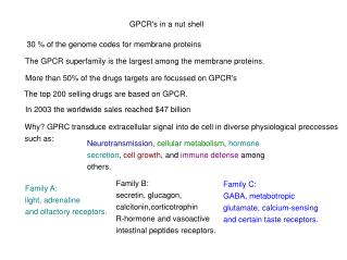
GPCR's in a nut shell 30 % of the genome codes for membrane proteins The GPCR superfamily is the largest among the membrane proteins. More than 50% of the drugs targets are focussed on GPCR's The top 200 selling drugs are based on GPCR. In 2003 the worldwide sales reached $47 billion Why? GPRC transduce extracellular signal into de cell in diverse physiological preccesses such as: Neurotransmission, cellular metabolism, hormone secretion, cell growth, and immune defense among others. Family B: secretin, glucagon, calcitonin,corticotrophin R-hormone and vasoactive intestinal peptides receptors. Family C: GABA, metabotropic glutamate, calcium-sensing and certain taste receptors. Family A: light, adrenaline and olfactory receptors.
Rhodopsin: opsin+retinal Function: Rh is involved in the molecular transformation of light into a neuronal signal. Structure 348 residues 7 TM helices + 1 cytoplasmic helix Motif DRY where D is forming double Salt bridge with E134 and E247 Important for maintaining the inactive states Extracellular loops (EL) well packed Intracellular loops (IL) coiled and with high B-factors. PDB: 1F88(2.8 A), 1U19(2.2 A) Disulfide bridge C187-C110 Palczewski K, et al. (2000) Science 289:739 2167 times cited
Vision Cycle: twist angles at C11-C12 (-18 ) C11 C12 + photon H20 + The torsion in the C11-C12 bond might be a pre-requisite for the isomerization process. Okada T. et al. (2004) JMB 342:571
Water network in rhodopsin NH+ of the Schiff base group at H-bond distance of COO- of Glu113. Thr94, Ser186 and Wat2b form a hydrogen bonding network involving Glu113. Wat2a extends the hydrogen bond to Glu181. The network continues up to Tyr268 and Tyr192. This network might a possible path for the switch of counterion in metarhodopsin I.
Squid rhodopsin 448 residues 7 TM helices + 2 cytoplasmic helices TM5 and TM6 show a 25 A extension into the cytosol.. TM6 interacts with C-terminus and Helix-9. The rigid conformation in the cytosol might be a structural motif associated binding to specific G-proteins. Disulphide bridge C186-C108
Helix-8 is anchored to the membrane by a palmitoyl group bonded to Cys337 Protein-protein interaction in the crystal structure is mediated by a phospholipid in the extracellular interface
The retinal is bonded to Lys 305 and shows a U shape around the Trp274 ring. There are five aromatic rings close to the retinal. The residues in contact with retinal show significant differences with respect to bovine rhodopsin. Glu180 is far away from retinal. Instead, Asn185 might mediate the interaction between retinal and Glu180 after photoisomerization.
Retinal environment in squid rhodopsin Retinal interactions Water network Larger amount of water molecules in the interhelical cavity than in bovine rhodopsin. Evidence of the change of vibrational frequencies of more than eight water molecules on formation of bathorhodopsin.
Final remarks New insights for the activation of membrane proteins can be addressed from the molecular dynamics simulation of squid rhodopsin. Light induce conformational changes in squid rhodopsin and the signal propagates towards the cytoplasmic side along the water cluster located in the interhelical domain.