Development of a Stem Cell-Based Pacemaker Utilizing Gene-Engineered Stem and Heart Cells
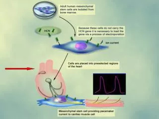
This study explores the hypothesis that a genetically engineered stem cell, when coupled with a heart cell, can function as a biological pacemaker. We aim to create a two-cell functional syncytium by utilizing gap junctions between stem cells and myocytes. Our methodology includes injecting quantum dot (QD) labeled heart mesenchymal stem cells (hMSCs) intramuscularly and at the epicardial border, along with tissue adhesive to promote adhesion. The study goals involve enumerating cell populations, performing 3D reconstructions, and characterizing the spatial distribution of quantum dots, visualized through histological sections using Hoechst 33342 for nuclei staining.

Development of a Stem Cell-Based Pacemaker Utilizing Gene-Engineered Stem and Heart Cells
E N D
Presentation Transcript
A C B D
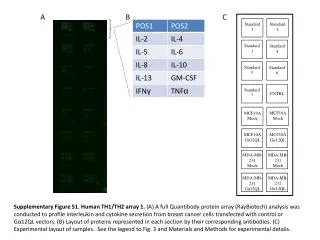
HCN Our basic premise: A Stem Cell Based Pacemaker requires a two cell functional syncytium Hypothesis: A genetically engineered stem cell coupled to a heart cell can serve as a biological pacemaker. Gap Junction Stem cell Myocyte
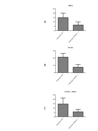
QD (red)+ Phase z 0 QD+hMSCs in needle track Intramuscular QD+hMSCs Epicardial border Tissue adhesive adsorbed to epicardium Imaging QD-hMSCs in histologic sections • Goals of study • Enumerate cells • 3-D reconstructions • Characterize distribution 1 mm Quantum dots Hoechst 33342 (nuclei)