Chapter 32: Leaf Structure and Function
Chapter 32: Leaf Structure and Function . Function – photosynthesis Shape – max. light absorption Diffusion of CO2 and O2 Ordered arrangement for light Loss of water vapor Trade off between photosynthesis and water conservation. External form.

Chapter 32: Leaf Structure and Function
E N D
Presentation Transcript
Function – photosynthesis • Shape – • max. light absorption • Diffusion of CO2 and O2 • Ordered arrangement for light • Loss of water vapor • Trade off between photosynthesis and water conservation
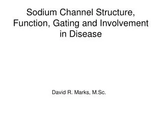
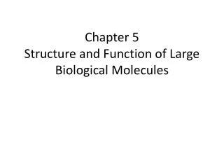
External form • Shapes – round, need, scalelike, cylindrical, heart, fan, thin, narrow • Size – 20m to < .5 cm • Blade, petiole, stipules • Simple, compound • Axil region
Fig. 35-6a (a) Simple leaf Petiole Axillary bud
Fig. 35-6b Leaflet (b) Compound leaf Petiole Axillary bud
Fig. 35-6c (c) Doubly compound leaf Leaflet Petiole Axillary bud
Fig. 35-6 (a) Simple leaf Petiole Axillary bud Leaflet (b) Compound leaf Petiole Axillary bud (c) Doubly compound leaf Leaflet Petiole Axillary bud
Leaf arrangement • Alternate – 1 leaf each node • Opposite – 2 leaves each node • Whorled – 3+ leaves each node
Leaf Venation • Veins = vascular tissue • Parallel • Netted • Palmately – from 1 point • Pinnately – branch from entire length of midvein
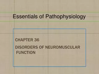
Leaf tissues • Upper epidermis + Lower epidermis • No chloroplasts/transparent • Cuticle – waxycutin • Trichomes – hairlike (fuzzy) • Retain moisture next to leaf, reflect light • Secrete irritants – herbivores • Texture – deter insects walk/eat • Excrete excess salts
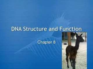
Fig. 35-9 EXPERIMENT Bald pod (no trichomes) Very hairy pod (10 trichomes/ mm2) Slightly hairy pod (2 trichomes/ mm2) RESULTS Very hairy pod: 10% damage Slightly hairy pod: 25% damage Bald pod: 40% damage
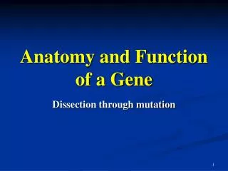
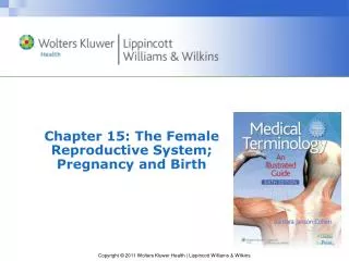
Subsidiary cells – epidermal; water and ions supplied to guard cells • Stomata (opening) + guard cells • Open/close stoma • Only epidermal cells with chloroplasts • Lower epidermis (land); upper epidermis (aquatic)
Fig. 35-18b Guard cells Stomatal pore 50 µm Epidermal cell (b) Surface view of a spiderwort (Tradescantia) leaf (LM)
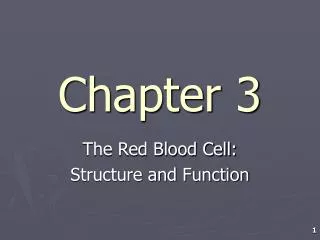
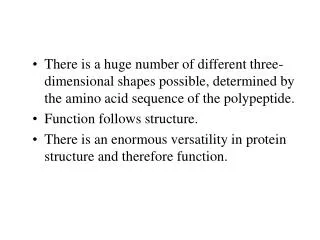
Mesophyll – photosynthetic ground tissue • Btw. Upper and lower epidermis • Parenchyma – chloroplasts • Air spaces – gas exchange • 2 sublayers: • Palisade mesophyll – top, columnar cells, close together • photosynthesis • Spongy mesophyll – lower, loose and irregularly shaped • Gas exchange
Vascular bundles – veins – through mesophyll • Xylem (top) and phloem (bottom) • Bundle sheath • Nonvascular, around vein • Parenchyma or sclerenchyma
Fig. 35-18 Guard cells Key to labels Stomatal pore 50 µm Dermal Epidermal cell Ground Cuticle Sclerenchyma fibers Vascular Stoma (b) Surface view of a spiderwort (Tradescantia) leaf (LM) Upper epidermis Palisade mesophyll Spongy mesophyll Bundle- sheath cell Lower epidermis 100 µm Cuticle Xylem Vein Phloem Vein Air spaces Guard cells Guard cells (a) Cutaway drawing of leaf tissues (c) Cross section of a lilac (Syringa)) leaf (LM)
Fig. 35-18a Key to labels Dermal Ground Cuticle Sclerenchyma fibers Vascular Stoma Upper epidermis Palisade mesophyll Spongy mesophyll Bundle- sheath cell Lower epidermis Cuticle Xylem Vein Phloem Guard cells (a) Cutaway drawing of leaf tissues
Fig. 35-18c Upper epidermis Key to labels Palisade mesophyll Dermal Ground Vascular Spongy mesophyll Lower epidermis 100 µm Air spaces Guard cells Vein (c) Cross section of a lilac (Syringa) leaf (LM)
Functioning of Stomata • Day – open – photosynthesis • Water moves into guard cells turgid + bend pore • Night – close • water leaves guard cells flaccid collapse close pore • Prolonged drought – stomata close (even in day) • Drop in CO2 in leaf – stomata open, even in dark • Photosynthesis (occurs in light) reduces internal concentration of CO2 in leaf, triggering stomata to stay open
Details of Stomatas Opening/Closing • H+ and K+ move across PM of guard cells • Blue light triggers K+ to move into guard from subsidiary/epidermal cells • Active transport – ATP • ATP provides energy to pump H+ out of guard • Removal of H+ makes electrochemical gradient to drive uptake of K+ • Uptake of K+ in guard increases solute conc. In vacuoles water enters guard from surrounding cells by osmosis
Result increase in turgidity changes guard shape • Almost opposite happens to close stomata • Evidence that increase in Ca2+ conc. In guard triggers closure
Transpiration • Loss of water vapor by evaporation • Responsible for water movement in plants • Factors influencing rate: • Temperature • Light • Wind + dry air
Benefits • Cools stems and leaves • Distributes minerals • Harmful effects • Loose more water than take in during heat loss of turgidity wilt • Temporary wilting of plant can “come back”
Leaf Abscission • Fall off, once/year • Many changes • Plant hormones – ethylene, abscisic acid (ABA) • Abscission zone – near base of petiole • Weak, parenchyma and few fibers
Modified leaves • Spines – animals • Tendrils – vine attachment • Bud scales – winter buds • Bulb – short underground stem with fleshy leaves for storage • Succulent leaves – water storage in dryness • Insectivorous plants