BACTERIA
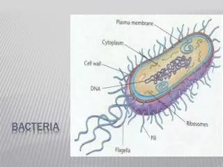
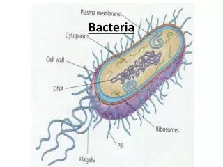
BACTERIA. CLS 311: Basic Bacteriology Mrs. Amany Ahmed Niazy. Bacterial Structure. Bacterial Structure. Exterior Structures. The exterior structure (cell envelope) is made up of two to three layers: I n some species of bacteria an outer capsule. C ell wall. C ytoplasmic membrane.

BACTERIA
E N D
Presentation Transcript
BACTERIA CLS 311: Basic Bacteriology Mrs. Amany Ahmed Niazy
Exterior Structures • The exterior structure (cell envelope) is made up of two to three layers: • In some species of bacteria an outer capsule. • Cell wall. • Cytoplasmic membrane.
Capsule • Some bacteria surround themselves with Capsule. • Most capsules are polysaccharides made of single or multiple types of sugar. • Capsule do not contribute to growth and multiplication. • Capsules provide some general protection for bacteria eg. Protect it from drying. . • Capsule major function in pathogenic bacteria is protection from the immune system (protect it from phagocytosis). The capsule is a major virulence factor in the major disease-causing bacteria, such asStreptococcus pneumoniae. (Noncapsulated mutants of these organisms are avirulent, i.e. they don't cause disease).
Bacterial capsules are non-ionic, so neither acidic nor basic stains will adhere to their surfaces. The medium in which the culture is grown as well as the temperature at which it is grown and the age of the culture will affect capsule formation
Cell Wall – Why is it important? • The rigid cell wall gives the bacterium its shape and surrounds the cytoplasmic membrane, protecting it from the environment. • The strength of the wall is responsible for keeping the cell from bursting when there are large differences in osmotic pressure between the cytoplasm and the environment. • It also helps to anchor appendages like the pili and flagella, which originate in the cytoplasmic membrane and protrude through the wall to the outside.
Structure of Cell Wall The cell wall of bacteria is composed of peptidoglycan, which covers the entire surface of the cell. It is made up of a combination of peptide bonds and carbohydrates (protein-sugar) • Peptidoglycan is a huge polymer of interlocking chains of identical monomers. The backbone of the peptidoglycan molecule is composed of two derivatives of glucose: N-acetylglucosamine (NAG) and N-acetlymuramic acid (NAM). The NAG and NAM strands are connected by interpeptide bridges.
Cell Wall • Several antibiotics (penicillins) stop bacterial infections by interfering with cell wall synthesis, while having no effects on human cells.
Classify pathogenic bacteria is on the basis of Gram Staining and Shape. Gram Stain Gram Positive Gram Negative Cocci Rods Cocci Rods
Structure of Cell Wall • The wall of a bacterium is classified in two ways: • Gram-positive. A gram-positive cell wall has many layers of peptidoglygan (up to 90% of the cell wall). Which makes it retain the crystal violet dye when the cell is stained. This gives the cell a purple color when seen under a microscope. The cell wall also contain Teichoic & lipoteichoic acids which promote adhesion and anchor wall to membrane. • Gram-negative. The cell walls of gram negative bacteria are more chemically complex. Peptidoglycan makes up only 5 – 20% of the cell wall, and is not the outermost layer, but lies between the plasma membrane and an outer membrane. This outer membrane is similar to the plasma membrane, but is less permeable and composed of lipopolysaccharides (LPS). LPS is a harmful substance classified as an endotoxin.
The Outer Membrane Of Gram Negative Bacteria. • Unique lipid by layer imbedded by protein. • It contain Porinsspecialized channel-forming proteins that can allow small molecules and ions to cross the outer membrane. • It is made up of lipid bilayer where the outside layer is made up oflipopolysaccharides (LPS) • Eg. Lipid A it is protein of LPS small amount of Lipid A stimulate immunity and defense system responds effectively and eliminate the invader. Lipid A large amounts example in GNR in blood stream the defense system damage even our won cells and this is the symptoms associated with endotoxin. • O-specific polysaccharide it differes among species of bacteria and it can be used to identify species or strains. Eg. E.coli O157:H7
Peptidoglycan and Antibiotics • Some antibiotics such as Penicillins and Cephalosporins, interfere with the linking of the interpeptides of peptidoglycan, but because of the LPS membrane, these antimicrobials can’t access the peptidoglycan of gram-negative bacteria. While gram-positive bacteria, are more susceptible to these antibiotics because of the lack of the LPS (lipopolysaccharides). Since the eukaryotic cells of humans do not have cell walls, our cells are not damaged by these drugs.
GRAM STAIN • There are two main types of bacterial cell walls, Gram positive and Gram negative, which are differentiated by their Gram staining characteristics. • Gram stain Procedure: • Crystal violet • Iodine • Alcohol • Saffranine
Gram +veandGram –veCell Wall • Gram Positive Color of Bacteria: Blue-violet • Gram Negative Color of Bacteria: RED
Cell Membrane (=cytoplasmic membrane) Cell membrane is composed of phospholipids bilayer and proteins which is found through out the living world. • It is responsible for selective and active transport of materials in and out of the cell. • It is involved in secretion of some exotoxins and hydrolytic enzymes involved in the pathogenesis of disease.
Cell Membrane (=cytoplasmic membrane) • It contain many proteins • Those proteins are constantly moving. • For example: more than 200 different membrane proteins have been found in E.coli. Many act as a receptores.
Cell Membrane Permeability • Simple Diffusion: • Water small hydrophobic molecules and gases. • Transport system: • Facilitated diffusion passive transport. • Moving in and out until their concentration is same on bothy side of the membrane. • It only eliminate difference in concentration but cannot create one. • Active transport move compounds against concentration gradient.
Filamentous Protein Appendages • Anchored in the cytoplasmic membrane and protrude out from the surface. • Not essential but help in survival.
Flagella, Appendages • Flagella (singular, flagellum) are long hair like protein structure that are found in many species of bacteria. • They may be found at either or both ends of a bacterium or all over its surface. • Function of the Flagella: the flagella beat in a propeller-like motion to help the bacterium move toward nutrients, or away from toxic chemicals.
Flagella, Appendages • It can rotate > 100,000 revolutions /min (move bacteria 20 body lengths/sec) • Eg. Helicobacter pylori multiple flagella at one end allow it to penetrate the viscous mucous gel that coats the stomach epithelium .
Structure of Flagella • Filament the portion extending into the exterior environment. It is composed of identical subunits of a protein called flagellin. Which form a helical structure with a hollow core. • Basal Body anchors the flagellum to the cell membrane. Gram negative contain 2 pairs of rings. Gram positive contain 1 pair of ring. • Hook connect the filament to the cell surface.
A) Tumbles last only a fraction of a second, which is sufficient to effectively randomize the direction of the next run. B) Runs tend to be variable in length extending from a fraction of a second to several minutes Cells tumble less frequently when they sense they are moving closer to an attractant. In contrast they tumble more frequently when they sense they are moving closer to a repellent.
Flagella, Appendages • Example of movements: • Chemotaxis bacteria moves toward a compound if it is a nutrient. • Phototaxis some bacteria respond to variations in light. • Aerotaxis bacteria respond to concentration fo oxygen.
Pili, Appendages • Pili(singular, pilus) are short hair-like projections found all around the surface of cells of many bacteria. • Composed of protein subunits arranged helically to form a long cylindrical molecule with a hollow core.
Function of the Pili • To enable attachment of cells to specific surfaces . It adhere by binding to a very specific molecule (called fimbriae). (e.g. E.coli that cause severe watery diarrhea attach to the cells that line the small intestine through specific interactions between pili and intestine surface). Without pili, many disease-causing bacteria lose their ability to infect because they are unable to attach to host tissue. • It play a role in movement of population of cells on solid media. • Some are called sex pilus because it is used to join one bacterium to another as a bridge for specific type of DNA transfer.
Cytosol (cytoplasm of the bacteria) • It is where the functions for cell growth, metabolism, and replication are carried out. • It is a gel-like matrix composed of water, enzymes, nutrients, wastes, and gases and contains cell structures such as ribosomes, a Nucleoid, and plasmids. • The cell envelope encases the cytoplasm and all its components. • Unlike the eukaryotic (true) cells, bacteria do not have a membrane enclosed nucleus.The chromosome, a single, continuous strand of DNA, is localized, but not contained, in a region of the cell called the nucleoid. All the other cellular components are scattered throughout the cytoplasm.
The Nucleoid The nucleoid is a region of cytoplasm where the chromosomal DNA (chromosome) is located. • The chromosome of prokaryotes is an irregular mass within the cytoplasm, that is usually attached to the cytoplasmic membrane. • It is usually a large, circular molecule of double-stranded DNA. It is usually tightly packed into about 10% of the total volume of the cell. • The absence of nuclear membrane is very important for rapid growth or prokaryotic cells in changing environments. Nucleoid
Ribosomes Ribosomes Involved in protein synthesis, they translate the genetic code form nucleic acid to that of amino acids • It is much more abundant than in the cytoplasm of eukaryotic cells this is a reflection of the higher growth rate of bacteria. • Ribosomes of prokaryotic cells 70S (composed of 2 subunits 30S – 50S) are smaller in size than ribosomes of eukaryotic cells 80S. • They differ in structure wich make them a target for certain antibiotics.
Storage Granules • Used to store nutrient that the cell has in relative excess. • Bacteria use granules to store minerals and nutrients (lipids, carbohydrates, phosphates, sulfur or metals) for the cell to use when needed.
Plasmids • Plasmids are small usually circular, double-stranded DNA. • It is separated from the chromosome, and they are not involved in reproduction. • They are found in many strains of bacteria. • A single bacterial cell can harbor multiple types of plasmids. Plasmids replicate independently of the chromosome and, while not essential for survival, appear to give bacteria a selective advantage. For example, many plasmids code for the production of one or more enzymes that destroy certain antibiotics (resistance to that antibiotic).
How are plasmids passed on from one bacteria to the other?? • Plasmids are passed-on to other bacteria through Two ways: 1. For most plasmid types, copies in the cytoplasm are passed on to daughter cells during binary fission.
How are plasmids passed on from one bacteria to the other?? • Other types of plasmids form a tube-like structure at the surface called a pilus that passes copies of the plasmid to other bacteria during conjugation, a process by which bacteria exchange genetic information.
Plasmids • Many plasmid genes promote survival and pathogenesis. • Plasmid are responsible for transfer of cellular properties such as, production of toxins, production of pili , resistance to antimicrobials and other toxic chemicals. • The ability to insert specific genes into plasmids have made them extremely useful tools in the fields of molecular biology and genetics, specifically in the area of genetic engineering.
Endospores • Endospores are bacterial survival structuresthat are highly resistantto many different types of chemical and environmental stresses and therefore enable the survival of bacteria in environments that would be lethal for these cells in their normal vegetative form. • Resistance of spore is due to dehydrated state, and specialized coats. • Germination of spores reproduces cell identical to that which was sporulated.
Endospores • Endospores may remain dormant for 100 years or even longer. Immersion in boiling water for hours may not kill them. Endospores that survive these treatments can germinate or exit the dormant stage, to become a typical multiplying cell, called a vegetative cell. • They can be found virtually anywhere. (cultivating media, soil, medical devices, food…etc) • Example of endo-spore forming bacteria • Clostridium botulinum • Clostridium tetani • Clostridium perfringens • Bacillus anthracis.
Spore Coat protein • Core Wall and Cortex peptidoglycan • Core metabolically inactive cell with low water content. When the environmental conditions are suitable, the endospore absorbs water, swells and the wall splits, releasing the cell inside. It develops a new cell wall and starts functioning as a typical bacterial cell.
Cell Morphology & Shape of Bacteria Coccus(spherical):e.g. Streptococci, Staphylococci Spirillum(spiral): e.g.Treponemaspp. Bacillus (rod-like): e.g.Enterobacteriaceaspp.