Download
1 / 41
450 likes | 535 Vues
Learn how CD169+ macrophages capture and present soluble antigens in lymph nodes, influencing the immune response to pathogens and tumors. Explore the process of antigen sampling and activation of B cells, essential for antitumor immunity.

E N D
CD169+ Macrophages, Lymph node sentinels. Rafael Eduardo Giuliano
Wholemicroorganisms and/orLargeantigens Exposure of uncommon antigens Capture oftheseantigens SOLUBLE ANTIGENS - VIA FLOW LYMPH = (LYMPH-BORNE) Migratorydendriticcells Lymphnode Presentationofantigens Adquired immune response
Nanometre log scale of pathogens ? ...how this presentation occurs Small antigens (<70kD) Complexed to antibodies Microbial antigens Solubleantigens Lipids Answer: Lymph node-resident APCs Dead cell-associated antigens (?) Bachmann & Jennings. 2010 Nat Rev Immuno MODIFIED
SecondaryLymphoidOrgan (SLO) _ BasicBuildingBlocks_ Junt et al. 2008 Nat Rev Immuno
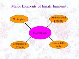
Antigen-sampling zone = Subcapsular sinus Subcapsular macrophages = Metallophilic macrophages = CD169+ macrophages von Andrian et al. 2003 Nat Rev Immuno
Subcapsularmacrophages = Metallophilicmacrophages = CD169+ macrophages Fibroblastic Reticular Cell Junt et al. 2008 Nat Rev Immuno
CD169+ macrophages as a FILTER Convencional resident DC (CD8+α) Small antigens (<70kD) Complexed to antibodies Microbial antigens Solubleantigens CD169+ macrophages Lipids Dead cell-associated antigens (?) Batista et al. 2009 Nat Rev Immuno
CD169+ = Siglec -1 = Sialoadhesin • Siglec (Sialic acid binding Ig-like Lectin-C) • Sialicacid: genericterm for a familyofnine-carbon • sugarsthat are derivativesofneuraminicacid • Involved in thedirectrecognitionof • sialylatedglycoconjugates. Crocker et al. 2001 Trends in Immuno
Expressionpattern ofhumanSiglecs Crocker et al. 2001 Trends in Immuno
...on the context of a tumor Defective antigen presentation (Dendritic cells) Immunosupressive environment induced by tumors Tolerance (T cells) Major source of tumor antigens: Dead tumor cells (apoptose) Anticancertherapy Dead cell-associated antigens (Soluble antigens) Lymph nodes CD169+ macrophages Presentation by cross-priming!!!!
Antitumorimmunity Dead cell-associated antigens (Soluble antigens) Dmitry Gabrilovich 2004 Nat Rev Immuno
Effectsofanticancertherapyon tumor cells Zitvogel et al. 2008 Nat Rev Immuno
‘Find-me’ and ‘eat-me’ signals and some phagocyte receptors Ravichandran et al. 2007 Nat Rev Immuno
Lymph nodes prevent the systemic dissemination of pathogens • Staging ground of adaptive immune responses; • How lymph borne virus particles are cleared from afferent lymph and presented to B cells ? Eduardo
How virus particles that enter peripheral tissues are handled within draining lymph nodes? Multiphoton Intravital Microscopy (MP-IVM) 0’ 30’ Popliteal Lymph Nodes UV -VSV Ultraviolet inactivated vesicular stomatitis virus VSV accumulates on the SCS floor
Which are the preferred VSV capturing cells in lymph nodes? WT - BM Act (EGFP) VSV are captured by hematopoietic cells Enhanced GFP in NON-hematopoietic cells
Which are the VSV capturing leukocytes? Electron Microscopy – Popliteal lymph nodes (5min after injection VSV) VSV selectively bound on the surface of large cells residing within the SCS or just below de SCS floor.
The VSV retaining cells belong to macrophages population? Confocal microscopy (30 min after injection UV-VSV) VSV accumulate rapidly and selectively on macrophages in the SCS of draining lymph nodes.
What are the consequences of viral capture by SCS macrophages for virus dissemination and antiviral immunity? *Viable VSV VSV titres 2h/6h after injection Untreated CLL treated Depletion of CD169+ macrophages rendered VSV filtration inefficient CLL Clodronate liposomes
How captured VSV is recognized by B cells? Electron Microscopy – Popliteal lymph nodes (30 min after injection VSV) Viral particles are presented to B cells within superficial follicles by macrophages that extend across the SCS floor
How the SCS macrophages influence the B cells distribution on draining lymph nodes? VSV-IND (Indiana Virus) VSV-NJ (New Jersey) (WT mice) WT B Cells *** + VI10YEN B Cells ** +/- CLL MP-IVM Confocal Microscopy Specific B Cells rapidly accumulated below and within the SCS floor VI10YEN B Cells VSV-IND (Indiana Virus)
What is the role of the SCS macrophages on the B cell activation? Surface IgMs on B cell populations on draining lymph node by flow citometry after VSV-IND injection No virus 30min 1h 2h No virus 2h UV-VSV CLL Treated Untreated
What is the role of the SCS macrophages on the B cell migration towards the T/B border? VSV-IND WT mice WT B Cells *** OR VI10YEN B Cells ** + /- CLL 6h Confocal micrograph Even without SCS macrophages, B cells are eventually activated by VSV-derived antigens, although less efficiently
Conclusion Capture of lymph borne viruses and guide them to presentation and activation of B cell
Aim: To identify how APCs in the lymph node (LN) internalize and crosspresent soluble antigens (dead tumor cells) to CD8+ T cells.
Immunization with dead tumor cells activates antitumor immunity? OT-I T cell - CFSE labeled OVA-expressing dead cell i.v. WT 64hr Proliferation FACS EG7 cells - Xray 10 days live EG7 cells / left flank tumor volumes s.c. /right flank WT ndLN dLN This immunization serves as na effective tumor “vaccination” by activation of specific CTL
What is the contribution of migratory DCs to the delivery of cellular antigens? Hind leg foot pads PBS or CFA or Dead Cell (apoptotic tumor cells) 12 -18hr 30hr CD11c+ DCs of dLN Violet light Kaede mice (Tg) Migratory DCs do not participate in the delivery of dead cells-associated antigens Kaede - photoconvertible fluorescence protein
How antigens reach the lymph node? Until 4 days 24hr PKH26- labeled dead cells (EG7) s.c. /foot pads 3, 6, 9, 18, hr and 4 days dLN Cell corpses traveled to the draining LNs shortly after the injection via lymphatic flow and were trapped in the sinus by CD169+ MΦ
Which cells phagocytosed the apoptotic cell corpses on dLN? 24hr PKH26-labeled dead cells (EG7) Sorting FACS (dLN) ndLN dLN Predominantly residents / non-migratory CD169+ MΦ
CD169+ MΦ depletion can change antitumor immunity? EG7-Xray WT day 14 EG7 live cells / left flank tumor volumes s.c. /right flank CD-169-DTR Mice depleted of CD169+ MΦ could no longer reject viable tumor cells
Tumor degradation in vivo might be controled by CD169+ MΦ? EG7 /right flank WT day 7 day 12 OVA oxaliplatin dLN IFN-γ ELISA CD-169-DTR Both vaccination with tumor cells killed ex vivo and by degradation of established tumor in vivo is controled by CD169+ MΦ
Are CD169+ MΦ required to antigen-specific T cell proliferation? WT CFSE-labeled OT-I T cell s.c. foot pads dead cell-OVA or CFA-OVA dLN proliferation i.v. CD-169-DTR CD169+ MΦ are essential for the crosspresentation of dead cell-associated antigens
What is the efficacy of vaccination in these mice? WT OT-I T cell 2 days EG7 live cells / left foot pads 14 days EG7-Xray / right flank dLN CD8+ T cell CD-169-DTR To a efficacy of vaccination and priming of T CD8+ cells, CD169+ MΦ are essentially necessary
How is the crosspresentation of APCs in vitro? WT 24hr dLN APCs (magnetic beads) OT-I T cells proliferation 42hr CD-169-DTR dead cell-OVA IFN-γ ELISA 72hr Dependent of CD169+ MΦ in the LN sinus
How is the crosspresentation of APCs in vitro? Dependent of CD11c+ CD169+ MΦ in the LN sinus
What is the localization of CD169+ MΦ? CD169+ localized in thesinus, CD11c+ in the T cellzone, CD169+ CD11c+ in the cortical andparacorticalsinus
Are CD11c+ and CD11c- CD169+ MΦ differents in function? CD11c+ CD169+ MΦ CD11c- CD169+ MΦ LPS CpG Two CD169+ MΦ subsets have distinct cytokine production profiles
What is the molecular mechanism of apoptotic cell phagocytosis by CD11c+ CD169+ MΦ? WT W3 dead cell-OVA PKH26 D89E or E1E2PT 24hr dLN APCs Citometry i.v. CFSE-labeled OT-I T cell dead cell-OVA with PBS, D89E or E1E2PT s.c. foot pads dLN proliferation CD169+ MΦ phagocytose dead cells in a PS-dependent manner D89E and E1E2PT milk fat globule-EGF-factor 8 (MFG-E8) mutants
Conclusion - Paper Highlights ► Dead tumor cells in periphery accumulate in the draining lymph node sinus; ► CD169+ macrophages phagocytose and crosspresent dead cell-associated antigens; ► CD169+ macrophage-depleted mice fail to crossprime tumor-specific CD8 T cells; ► CD169+ macrophages link tumor cell death and induction of antitumor immunity.
Conclusion - Journal TUMOR INFECTION “Soluble” antigens are drained to lymph nodes CD169+ Macrophages sinus take this antigens and generate a humoral and cellular immune response!