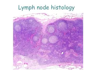
Lymph Node Normal Morphology
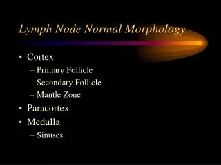
Lymph Node Normal Morphology. Cortex Primary Follicle Secondary Follicle Mantle Zone Paracortex Medulla Sinuses. Cortex. Primary B-Cell Follicles Nodules of small lymphocytes Lack germinal centers Secondary B-Cell Follicles Result of stimulation Germinal Centers Mantle zone.

Lymph Node Normal Morphology
E N D
Presentation Transcript
Lymph Node Normal Morphology • Cortex • Primary Follicle • Secondary Follicle • Mantle Zone • Paracortex • Medulla • Sinuses
Cortex • Primary B-Cell Follicles • Nodules of small lymphocytes • Lack germinal centers • Secondary B-Cell Follicles • Result of stimulation • Germinal Centers • Mantle zone
Germinal Centers • Pale zone • toward antigen entry • small cleaved cells / centrocytes • follicular dendritic cells • Dark zone • toward paracortex • large lymphoid cells / centroblasts • tingible body macrophages
Mantle Zone • polarized toward antigen entry • express bcl-2 protein
Paracortex • rich in T cells • CD4:CD8 ratio variable • interdigitating dendritic cell • S-100 positive • irregular vesicular nuclei • high endothelial venules • postcapillary vessel • cuboidal epithelium
Medullary areas • B cells predominate especially plasma cells • histiocytes
Handling the Fresh Specimen • Surgeon should excise the largest and most abnormal node • Tissue for histology • Touch imprints • Fresh / frozen tissue for immunologic studies • Sterile portion for cytogenetics
Frozen Section • Diagnostic frozen section should be discouraged • Use frozen to assess adequacy or triage tissue
Freezing for Immunologic Studies • Liquid nitrogen or isopentane / dry ice mix is best • Thin sections (<2 mm )may be frozen in OCT • OCT must be wrapped in foil / plastic to avoid desiccation • Store at -70°C ideal but -20°C suitable for many antigens
Fixation • Node sliced in 2-3 mm intervals • One metal based fixative (B5, Zenkers, zinc sulfate) • One neutral buffered formaldehyde (formalin)
Processing • Single most important factor for optimal histology is section thickness • Sections should be one cell layer thick
Routine Stains • H&E • Giemsa - highlight nuclear features, cytoplasmic granules and plasmacytoid features • PAS - highlights mucin and glycogen, immunoglobulin inclusions and blood vessels • Methyl-green pyronin - highlights plasmacytoid features
Common Errors in Fixation and Processing • Drying of specimen - dark edge artifact; autolysis if prolonged • Section >3 mm thick - soft unfixed core; center cells show ballooning and are pale • Overfixation in B5 - brittle tissue; decreased nuclear staining • Inadequate dehydration - numerous cracks (dry earth look)
Common Errors in Fixation and Processing • Paraffin too hot - muddy staining with poor detail • Improper sectioning - Venetian-blind effect; poor cytologic detail • Section drying too hot - bubbled nuclei and antigen loss