CH 20 Lymph Node Anatomy
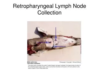
CH 20 Lymph Node Anatomy. James F. Thompson, Ph.D. The Lymph Nodes. Anatomy oval, bean shaped structures scattered throughout body along lymph vessels may be deep or superficial concentrated along the respiratory tree and GI tract, in the mammary glands, axillae, and groin

CH 20 Lymph Node Anatomy
E N D
Presentation Transcript
CH 20Lymph Node Anatomy James F. Thompson, Ph.D.
The Lymph Nodes • Anatomy • oval, bean shaped structures scattered throughout body along lymph vessels • may be deep or superficial • concentrated along the respiratory tree and GI tract, in the mammary glands, axillae, and groin • filter lymph fluid to trap foreign organisms, cell debris, and tumor cells
Lymphatic Organs – Lymph Nodes • Covered by a fibrous connective tissue capsule • Trabeculae extend from cortex to medulla • Stroma – the internal supportive connective tissue network of reticular fibers
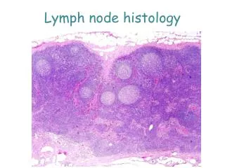
Structure of a Lymph Node • outer cortex - filled with lymph follicles • outer edge of follicle contains more T cells • inner germinal center is the site of B-cell proliferation • inner medulla - medullary cords of lymphocytes, macrophages, plasma cells (activated B cells) Cortex Medulla
Structure of a Lymph Node • Medullary cords extend from the cortex and contain B cells, T cells, and plasma cells • Throughout the node are lymph sinuses crisscrossed by reticular fibers • Macrophages reside on these fibers where they phagocytize foreign matter
Histology of Lymph Nodes follicles with germinal centers
Circulation in the Lymph Nodes • Lymph enters via a number of afferent lymphatic vessels • It then enters a large subcapsular sinus and travels into a number of smaller sinuses • It meanders through these sinuses and exits the node at the hilus via efferent vessels • The node acts as a “settling tank,” because there are fewer efferent vessels, lymph stagnates somewhat in the node • This allows lymphocytes and macrophages time to carry out their protective functions Only lymph nodes filter lymph!
Lymph Flow Through Lymph Nodes • fluid enters cortex through afferent vessels • filter and trap damaged cells, microorganisms, foreign substances, tumor cells by reticular fibers • macrophages phagocytize some, lymphocytes destroy some by immune defenses • exits medulla by efferent vessels at hilus
Blood Flow Through Lymph Nodes • blood vessels enter and exit at the hilus • this blood circulation provides nutrition for the node’s tissues and a route for leukocytes to enter into or exit from the lymphatic tissue of the node