Download
1 / 48
490 likes | 933 Vues
Normal Bone Growth and the Pathogenesis of Osteoporosis. Outline. Basic Skeletal Structures & Functions Mechanisms of Cellular Genesis Pathogenesis of Osteoporosis Pharmacological Therapy for Osteoporosis. Osteoporosis.

E N D
Outline • Basic Skeletal Structures & Functions • Mechanisms of Cellular Genesis • Pathogenesis of Osteoporosis • Pharmacological Therapy for Osteoporosis
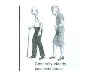
Osteoporosis Reduction in the quantity of bone or atrophy of skeletal tissues; occurs in postmenopausal women and elderly men, resulting in bone trabeculae that are scanty, thin and without osteoclastic resorption
Begin bone loss in their 40s Rapid loss of bone mass for 5-10 years after menopause onset Smaller bone cortices and diameter from growth phase, especially during puberty Begin bone loss in their 40s Bone loss remains linear and slow as sex steroid production progressively declines Larger bone cortices and diameter from growth phase Women vs. Men
Other Risk Factors • Chronic glucocorticoid excess • Hyperthyroidism • Inappropriately high T4 replacement • Alcoholism • Prolonged immobilization • Gastrointestinal disorders • Hypercalciuria • Malignancies • Cigarette smoking
Cells, Connective Tissue and Spaces • Highly specialized cells • Mineralized connective tissue matrix • Unmineralized connective tissue matrix • Spaces: bone marrow cavity vascular canals canaliculi lacunae
Modeling- During growth phase, the removal of bone at one site and desposition to another site Purpose- Achieve skeletal size and shape Remodeling- Once mature, a periodic replacement of old bone with new at the same location Purpose- Repair fatigue damage and prevent excess aging on load bearing bones Development/Growth
BMU MOVEMENT
Vital Statistics • Lifespan of BMU ~ 6-9 months • Speed ~ 25 mm/day • Bone volume replaced ~ 0.025 mm3 by a single BMU • Lifespan of osteoclast ~ 2 weeks • Lifespan of osteoblast (active) ~ 3 months • Interval between successive ~ 2-5 years remodeling events at the same location • Rate of turnover of whole ~ 10% per yr. skeleton
Osteoblastogenesis • Multipotent mesenchymal stem cell precursors • Reach bone by migration from neighboring connective tissue • Initiated by bone morphogenenetic proteins (BMP) • BMP activates core binding factor a1 (Cbfa1)
Osteoblastogenesis • Cbfa1 activates osteoblast genes: osteopontin, bone sialoprotein type 1 collagen, osteocalcin • BMP also stimulates distal-less 5 (Dlx5) • Dlx5 regulates osteocalcin, alkaline phosphatase and mineralization • Osteoblast differentiation stimulated by: TGFb, PDGF, FGF and IGF
Osteoblasts • Produce and secrete proteins for bone matrix • Also produce osteocalcin and osteonectin • Mineralization, the desposition of hydroxyapatite • Matrix synthesis = bone volume • Mineralization = bone density
Osteocytes • Osteoblasts buried in lacunae of matrix • Communicate inter- and extracellularly by extensions of the plasma membrane • Mechanosensory cells for bone growth or reduction and repair of microdamage • Sense changes in interstitial fluid and detect changes in hormone levels
Lining Cells • Osteoblasts that have become flat and elongated cells on the bone surface over a layer of unmineralized collagen • Possible functions: secrete collagenase produce homing signal for osteoclast precursors by direction of osteocytes
Osteoclastogenesis • Hematopoietic cell precursor • Reach the bone by circulation • Cytokines and colony-stimulating factors: IL-1, IL-3, IL-6, IL-11, LIF, OSM, TNF, GM-CSF, M-CSF, c-kit ligand • Inhibiting cytokines: IL-4, IL-10, IL-18, INF-g • Stimulating hormones- PTH and 1,25-dihydroxyvitamin D3
Interleukin-6 • Produced by cells of the osteoblastic lineage • Stimulates osteoclastogenesis and bone resorption • Binding recruits gp130, which initiates a tyrosine phosphorylation pathway • Also effects the differentiation of osteoblasts when in the presence of an IL-6 receptor
Interleukin-6 • Requires IL-6 membrane-bound receptors in order to begin osteoclastogenesis • Can initiate osteoclastogenesis in the absence of IL-6R if exogenous dexamethasone is added
Interleukin-6 • Osteoblastic stromal cells express very little IL-6R mRNA without dexamethasone treatment • Splenic and bone marrow cells do not require dexamethasone in order to express IL-6R mRNA • IL-11 does not require dexamethasone treatment to express IL-11R mRNA
Interleukin-6 • Requires pretreatment with dexamethasone in order to initiate a tyrosine phosphorylation pathway using gp130 signal transducing protein of osteoblastic cells
Interleukin-6 • Osteoclasts can be formed when human IL-6 is added to a coculture of splenic cells from a normal mouse and osteoblastic cells from a transgenic mouse overexpressing human IL-6R but not from a coculture of splenic cells from a transgenic mouse overexpressing human IL-6R and osteoblastic cells from a normal mouse
Conclusion • gp130 needs to be stimulated in order to produce osteoclasts • Cytokine-specific receptors are required to initiate a response to the cytokine • Dexamethasone allowed IL-6 to bind and initiate differentiation in osteoclasts in the absence of the receptor
Osteoclasts • Ruffled border erosion • Clear zone of suitable microenvironment • Degrade bone matrix • Endocytose matrix components and pass them through the cell to be exocytosed into the bone membrane
Apoptosis • 100% of osteoclasts die vs. 65% of osteoblasts • Osteocytes also undergo apoptosis, though they have longer lifespans than osteoclasts and osteoblasts • Growth factors and cytokines can initiate cell apoptosis: for example, TGFb stimulates osteoclastic apoptosis but inhibits osteoblastic apoptosis
Sex Steroid Deficiency • Up-regulating the production and action of cytokines that are responsible for osteoclastogenesis and osteoblastogenesis due to a loss of sex steroids results in up-regulation of osteoclast and osteoblast formation in the marrow • Stimulation of osteoclastogenesis exceeds that of osteoblastogenesis and results in bone loss
Estrogen Deficiency • Specifically, normal levels of estrogen suppress IL-6 activity with control of both IL-6 and IL-6R genes • Increase in bone resorption is due to increased osteoclastogenesis • Does osteoblastogenesis increase as well?
Ovariectomized Mice • Osteoblastic progenitors (CFU-F and CFU-OB) increase as a result of ovary removal • Larger colonies of osteoblastic precursors are less differentiated than smaller colonies • Smaller colonies are more committed to osteoblastogenesis
Ovariectomized Mice • Does ovary removal have an effect on progenitor proliferation? • What is the effect of ovariectomy on the expression of alkaline phosphatase?
Ovariectomized Mice • Initial volume of CFU-F increases and gradually falls to the control level • CFU-F and CFU-OB react in the same prolific manner, though there are many more CFU-F • Osteocalcin serum level initially increases and gradually falls to the control level
Ovariectomized Mice • Osteoclasts and osteoclastic precursors also initially increase but rapidly fall to the control level eariler after ovary removal than osteoblasts • Bone volume decreased more in ovariectomized mice than in the control group due to a 3-4 fold increase in osteoclastogenesis
Ovariectomized Mice • Is osteoblastogenesis dependent on factors released from the bone matrix during resorption?
Pharmacotherapy • A promising drug therapy for osteoporosis would be an anabolic drug that increases skeletal mass through rebuilding • PTH does increase bone mass, but is the mechanism by stimulation of osteoblast differentiation or prevention of osteoblast apoptosis?
Pharmacotherapy • PTH stimulated an increase of bone mineral density (BMD) in both normal mice and mice with osteopenia • The increase in the two strains occurred at an equal interval from their respective base-line values
Pharmacotherapy • Is the action of PTH directly toward the osteoblasts and osteocytes or indirectly by other secondary actions?
References • Jilka, RL, Takahashi, K, Munshi, M, Williams, DC, Roberson, PK, Manolagas SC. 1998. Loss of Estrogen Upregulates Osteobastogenesis in the Murine Bone Marrow. Journal of Clinical Investigation 101(5): 1942-1950. • Jilka, RL, Weinstein, RS, Bellido, T, Roberson, P, Parfitt, AM, Manolagas, SC. 1999. Increased Bone Formation by Prevention of Osteoblast Apoptosis with Parathyroid Hormone. Journal of Clinical Investigation 104(4): 439-446. • Manolagas, SC. 2000. Birth and Death of Bone Cells: Basic Regulatory Mechanisms and Implications for the Pathogenesis and Treatment of Osteoporosis. Endocrine Reviews 21(2): 115-137 • Roodman, GD. 1996. Advances in Bone Biology: the osteoclast. Endocrine Reviews 17(4): 66-80. • Udagawa, N, Takahashi, N, Katagiri, T, Tamura, T, Wada, S, Findlay, DM, Martin, TJ, Hirota, H, Taga, T, Kishimoto, T, Suda, T. 1995. Interleukin-6 Induction of Osteoclast Differentiation Depends on IL-6 Receptors Expressed on Osteoblastic Cells But Not on Osteoclast Progenitors. Journal of Experimental Medicine 182(11): 1461-1468.