Download
1 / 20
200 likes | 502 Vues
SEPARATION AND DETECTION OF PROTEINS part II Vlasta Němcová, Michael Jelínek, Jan Šrámek. SDS-PAGE ( = s odium d odecyl s u l phate- p oly a crylamide g el e lectrophoresis) - met hod for separation of protein s according to their size ( molecular weight ). Protocol:.

E N D
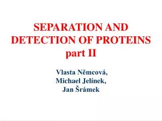
SEPARATION AND DETECTION OF PROTEINS part II Vlasta Němcová,Michael Jelínek,Jan Šrámek
SDS-PAGE(=sodium dodecylsulphate-polyacrylamide gel electrophoresis)- methodfor separation of proteinsaccording to their size (molecular weight)
Protocol: • preparation of polyacrylamide gels • mixing of tissue lysates with sample buffer • heating of samples at 95C • loading of samples onto polyacrylamide gel • electrophoresis of samples • staining of the gel with separated proteins in Coomassie Blue stain • destainingof the gel • localization of actin and myosin in the gel, comparison of their expression in tissues used • chemiluminiscent detection of actin
Separated proteins in polyacrylamide gel 1 2 3 4 5 6 7 --- Myosin (heavy chain) --- Actin --- Tropomyosin --- Myosin (light chain) --- Myosin (light chain) --- Myosin (light chain) 1. Marker 5. Actin 2. Liver 6. Myosin 3. Heart 7. Marker 4. Muscle
What is in sample buffer for SDS-PAGE? • Tris buffer provides appropriate pH • SDS (sodium dodecyl sulfate)- detergent that dissolves proteins and gives them a negative charge • glycerolmakes samples sink into wells during loading onto gel • bromophenol blue is tracking dye moving ahead of proteins – indicator of separation progress
Levels of protein organization • Primary structure = linear chain of amino acids • Secondary structure = domains of repeating structures, such as β-pleated sheets and α-helices • Tertiary structure = 3-dimensional shape of a folded polypeptide, maintained by disulfide bonds, electrostatic interactions, hydrophobic effects • Quarternaty structure = several polypeptide chains associated together to form a functional protein
Preaparation of samples for SDS-PAGE • before separation proteins have to be denaturated • denaturation by heating at 95°C in sample buffer containing SDS the proteins no longer have any secondary, tertiary or quaternary structure
Preaparation of samples for SDS-PAGE • resultant SDS-coated proteins take on a rod-like shape and a uniform negative charge-to-mass ratio proportional to their molecular weights speed of protein migration in gel depends ONLY on their molecular weight
How does an SDS-PAGE separation work? • negatively charged proteins move to positive electrode • smaller proteins move faster • proteins are separated by their size (molecular weight) _ +
Why use polyakrylamide gels to separate proteins? • smaller pore size than agarose • proteins are much smaller than chromosomes and routinelly separated fragments of DNA produced in PCR reaction • polymerization of acrylamide and N´,N´-methylenbisacrylamide is started by reaction initiator, amonium persulfate (APS), and a catalyst, tetramethylenediamine (TEMED)
Protein size (molecular weight) • measured indaltons (Da) or kilodaltons (kDa),1 kDa = 1000 Da • Dalton = atomic mass unit = 1 Da correspond to mass of hydrogen molecule (1.66 x 10 -24 g) • average nucleotide pair = 649 Da • average amino acid = 110 Da 200 AA protein – cca 20 kDa vs. 100 bp fragment DNA – cca 65 kDa
Muscles Contain Proteins of Many Sizes ProteinkDaFunction titin 3000 center myosin in sarcomere dystrophin 400 anchoring to plasma membrane filamin 270 cross-link filaments into gel myosin heavy chain210slide filaments spectrin 265 attach filaments to plasma membrane nebulin 107 regulate actin assembly a-actinin 100 bundle filaments gelosin 90 fragment filaments fimbrin 68 bundle filaments actin42form filaments tropomyosin 35 strengthen filaments myosin light chain27slide filaments troponin (T, I, C) 30, 19, 17 mediate regulation of contraction thymosin 5 sequester actin monomers
Separated proteins in polyacrylamide gel 1 2 3 4 5 6 7 --- Myosin (heavy chain) --- Actin --- Tropomyosin --- Myosin (light chain) --- Myosin (light chain) --- Myosin (light chain) 1. Marker 5. Actin 2. Liver 6. Myosin 3. Heart 7. Marker 4. Muscle
Western blot analysis(imunochemical detection of proteins) • transfer of separated proteins from the gel onto a membrane • identification of particular protein by imunodetection (=binding of primary and secondary antibody) • visualization by color reaction or chemiluminescence • the name of the method is a pun of the name SOUTHERN blot, a technique for DNA detection developed earlier by Edward Southern • similarly is named NORTHERN blot, technique for detection of RNA
Chemiluminiscent detection of actin equal volume of samples obtained form separate isolations heart heart heart MW muscle liver heart actin+myosin 30 ug of protein blot of SDS-PAGE gel
Examples of applications of Western blot in medicine • detection of antibodies (e.g. for confirmation of diagnosis) • boreliosis, EBV, HIV, HSV, Helicobacter pylori • autoantibodies, antibodies against nuclear antigens (ANA), antibodies against neural antigens
Principle of tests used in clinical praxis • Membrane strips containing electrophoretically separated antigen extracts are used as solid phase. The position of the proteins depends on their respective molecular masses. . • If the sample is positive, specific antibodies in the diluted serum sample attach to the antigens coupled to the membrane. • The attached antibodies react with AP-labelled anti-human antibodies (AP = alkaline phosphatase).
Principle of tests used in clinical praxis • The bound antibodies are stained with a chromogen/substrate solution which is capable of promoting a color reaction. An intense dark band at the line of the corresponding antigen appears if the serum sample contains specific antibodies. • Evaluating the band patterns on the incubated membrane strips involves differentiating non-specific from specific antibodies. The number and intensity of the specific bands is decisive for the result "positive/negative".
EUROLINE-WB: detection of antibodies against Borrelia EUROLINE ANA-Profile 3 Line immunoassay for confirmation and discrimination of antibodies HIV-1 and HIV-2