Download
1 / 31
310 likes | 435 Vues
This document details innovative methods of MRI segmentation applied to a patient with Secondary Progressive Multiple Sclerosis (SPMS). Utilizing an inverse nonlinear warp from MNI to SPGR FA and MPRAGE spaces, significant white matter demyelination and lesion identification was achieved, despite some limitations with z-score thresholding. Various segmentation techniques (SPGR, FLAIR, T2-PD) were assessed for their effectiveness in capturing cerebrospinal fluid (CSF), grey matter (GM), and white matter (WM) lesions. Comprehensive analysis highlights best practices and areas for improvement.

E N D
Notes • This was accomplished by doing an inverse nonlinear warp from MNI to the SPGR FA 18, then to the MPRAGE space with a linear transform (ideally these transforms should be combined) • Images are larger than shown, zoom in for more detail • Not all lesions are found by the z-score thresholding but a significant amount are • Large volumes of white matter are found to be significantly demyelinated: perhaps DAWM or actually revealing the “invisible disease”? • This was one the patients with a larger amount of low MWF volume, EDSS 5.0 SPMS
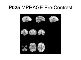
MPRAGE • Same slices as shown in the z-score slides • Lesions of interest circled
SPGR 3 class-CSF • Generally good CSF segmentation • Does not capture most lesions
SPGR 3 class-GM • Includes some or all of lesions
SPGR 3 class-WM • Matches nicely with the MPRAGE scan • Partially includes lesions
SPGR 4 class-CSF • Generally worse than 3 class CSF
SPGR 4 class-GM • More conservative estimate of GM, much fewer lesions included
SPGR 4 class-More GM • Deep GM • Includes many of the lesions
SPGR 4 class-WM • Does well at excluding most focal lesions but appears to be some partially including some • More conservative
SPGR-FLAIR 3 class-CSF • Grabs most lesions • Unfortunately mis-classifies GM too
SPGR-FLAIR 3 class-GM • Includes many regions previously seen as WM
SPGR-FLAIR 3 class-WM • Good exclusion of lesions identified by z-score • Does poorly at WM and GM segmentation, misses some WM
SPGR-FLAIR 4 class-CSF • Generally worse than 3 class CSF also • Chokes back mask too far
SPGR-FLAIR 4 class-GM • Still misidentifies a lot of WM as GM • Catches edges of our lesions of interest
SPGR-FLAIR 4 class-More GM • Again mis-includes a lot of WM
SPGR-FLAIR 4 class-WM • Avoids lesions but also misses a lot of regular WM since those are misidentified as GM
SPGR-T2-PD 3 class-CSF • Decent at outside brain CSF, though catches some WM • Does not get any CSF inside brain
SPGR-T2-PD 3 class-GM • Captures our lesions of interest • Gets ventricle CSF • Not as good as SPGR 3 class
SPGR-T2-PD 3 class-WM • Overly greedy WM • Misses focal lesions but gets GM
SPGR-T2-PD 4 class-CSF • Same problems as with SPGR-T2-PD CSF segmentation
SPGR-T2-PD 4 class-GM • GM + inner CSF • Catches darker lesions • Pretty poor, also gets WM
SPGR-T2-PD 4 class-More GM • Catches some lesions but overall pretty garbagey • Doesn’t really correspond to a distinct tissue class
SPGR-T2-PD 4 class-WM • A conservative estimate • Circled region shows possible inclusion of GM and missing of brain stem
Best CSF • SPGR 3 class (includes some lesions) • SPGR-FLAIR 3 class (includes all lesions) • SPGR-T2-PD 3 class (useful for out of brain CSF, does not include lesions)
Best WM • SPGR 3 class (best anatomically) • SPGR 4 class (conservative) • SPGR-FLAIR 4 class (more lesion exclusion)
Best GM • SPGR 3 class (GM+lesions) • SPGR 4 class (deep GM+lesions)
Best Lesion • SPGR 3 class (GM+lesions) • SPGR-FLAIR 3 class (CSF missing some lesions)
Coming Soon • Segmentation with a priori maps • Ideas about how to combine maps to produce NAWM, NAGM, and lesion only masks